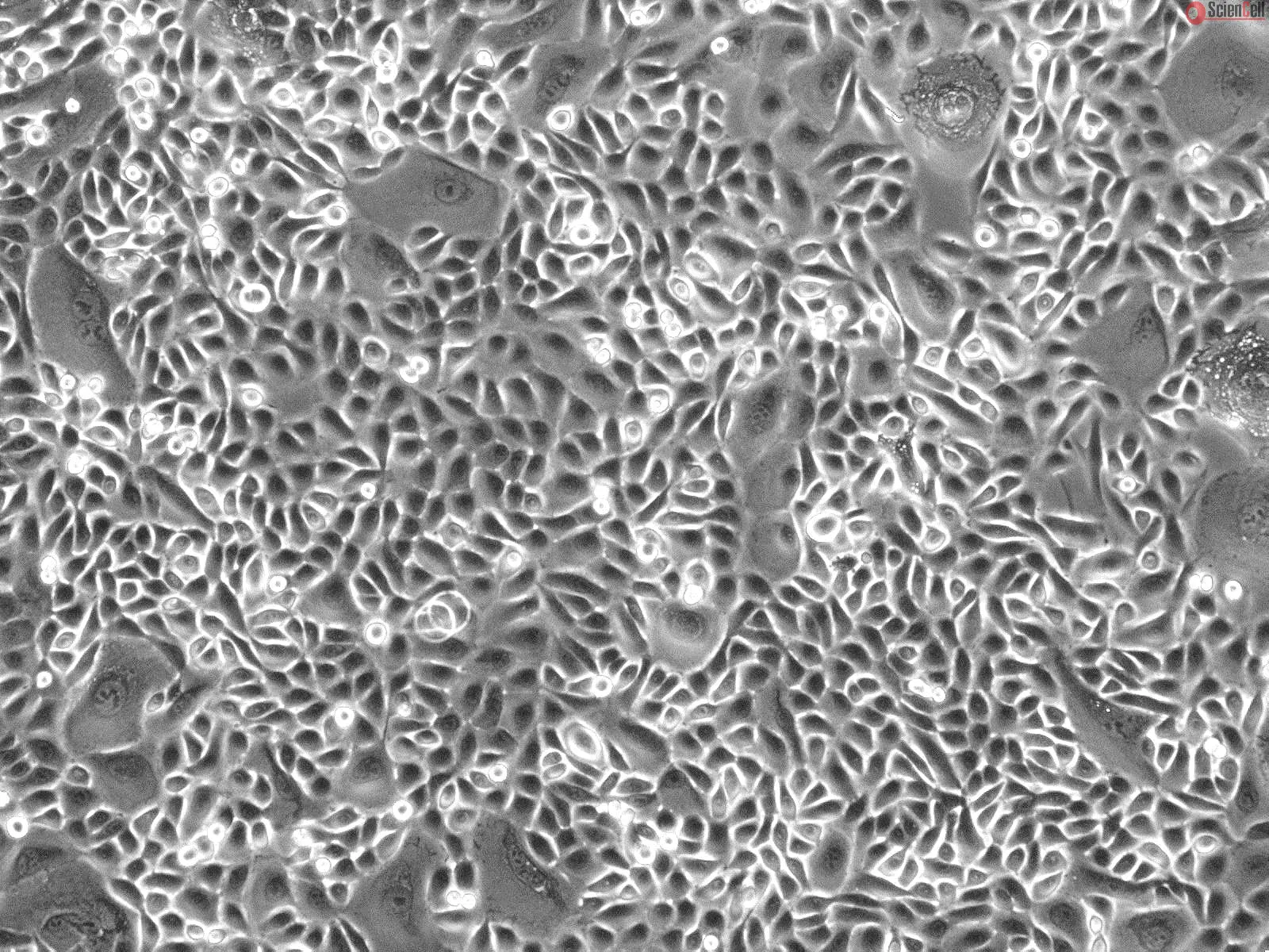
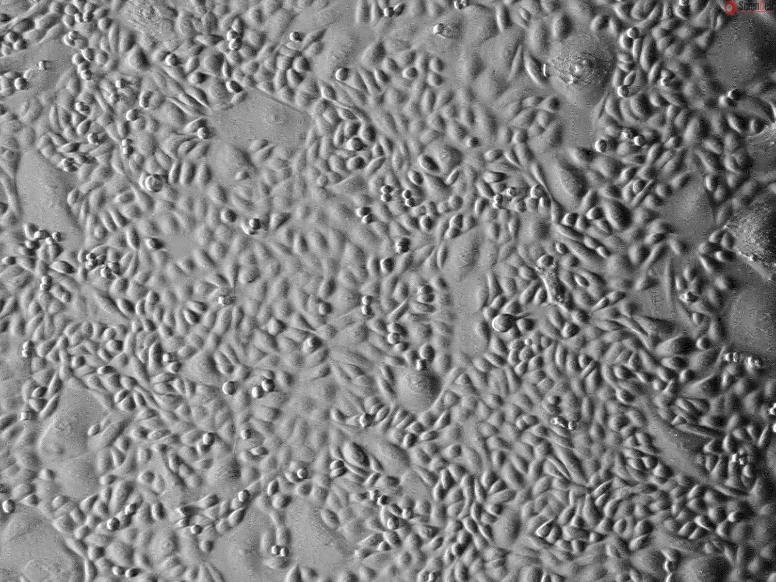
The epidermal layer of the skin provides an essential function as a protective barrier against insults from the external environment. The predominant cell type in the epidermis is keratinocytes and they are located in the stratified squamous epithelia. Keratinocytes are named after keratin, which is the most abundant protein in this cell. Progenitors of keratinocytes reside and divide in the basal layer of the epidermis. They then differentiate, migrate towards the surface of epidermis, and eventually withdraw from the cell cycle permanently. Keratinocyte proliferation, differentiation, and programmed cell death are complex and carefully choreographed processes. Apart from their protective functions, keratinocytes express adhesion molecules and cytokines, further suggesting an implication in skin innate immunity, tissue homeostasis, wound healing, cancer development, and skin-based gene-therapy.
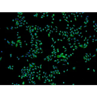
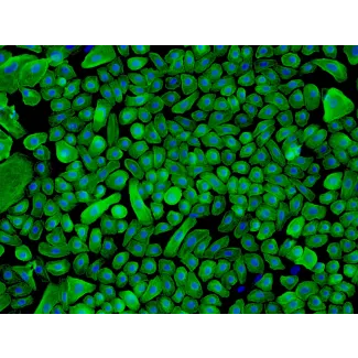
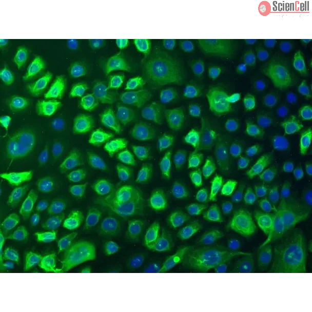
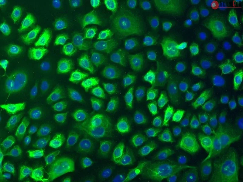
HEK-a from ScienCell Research Laboratories are isolated from adult human skin. HEK-a are cryopreserved at passage one and delivered frozen. Each vial contains >5 x 10⁵ cells in 1 ml volume. HEK-a are characterized by immunofluorescence with antibodies specific to cytokeratin-18 and/or cytokeratin-19. HEK-a are negative for HIV-1, HBV, HCV, mycoplasma, bacteria, yeast, and fungi. HEK-a are guaranteed to further expand for 10 population doublings under the conditions provided by ScienCell Research Laboratories.
Recommended Medium
It is recommended to use Keratinocyte Medium (KM, Cat. #2101) for culturing HEK-a in vitro.
Background Benvitimod (Tapinarof), as a small-molecule topical therapeutical aryl hydrocarbon receptor (AHR)-modulating agent, is in clinical development for treating pso... More
Background Benvitimod (Tapinarof), as a small-molecule topical therapeutical aryl hydrocarbon receptor (AHR)-modulating agent, is in clinical development for treating psoriasis and atopic dermatitis. Benvitimod reduces proinflammatory cytokines in psoriasis by specifically binding and activation of AHR. However, whether benvitimod can inhibit keratinocyte proliferation remains unclear. Minichromosome maintenance protein 6 (MCM6) is a key element of the prereplication complex (pre-RC) assembly which is one of the essential steps in the initiation of DNA replication for cell proliferation. Objectives This study aimed to determine whether benvitimod could reduce the excessive proliferation of psoriatic keratinocytes by inhibiting MCM6. Methods We examined the inhibitory effect of benvitimod on MCM6-mediated proliferation of keratinocytes by HaCaT cells in vitro and an IMQ-induced psoriatic model of mice in vivo. Results Epidermal MCM6 expression was enhanced in the skin lesions of psoriatic patients. The experiments further revealed that MCM6 was required for the proliferation of keratinocytes and governed by the IL-22/STAT3 pathway. In addition, the antiproliferation effect of benvitimod is achieved by the inhibition of p-JAK1 and p-JAK2, which further restrained the activation of STAT3 in keratinocytes. Lastly, benvitimod could repressed imiquimod-induced skin lesions and the expression of epidermal MCM6 and p-STAT3 in mice. Moreover, knockdown of AHR in keratinocytes enhanced the activation of JAK1 and JAK2. Conclusion The findings reveal that benvitimod could decrease MCM6-mediated proliferation of keratinocytes by affecting the JAK/STAT3 pathway, thereby serving as a new treatment modality for psoriasis. Less
We present a method to create multi-layered engineered tissue composites consisting of human skin fibroblasts and keratinocytes which mimic skin layers. Three-dimensional... More
We present a method to create multi-layered engineered tissue composites consisting of human skin fibroblasts and keratinocytes which mimic skin layers. Three-dimensional (3D) freeform fabrication (FF) technique, based on direct cell dispensing, was implemented using a robotic platform that prints collagen hydrogel precursor, fibroblasts and keratinocytes. A printed layer of cell-containing collagen was crosslinked by coating the layer with nebulized aqueous sodium bicarbonate. The process was repeated in layer-by-layer fashion on a planar tissue culture dish, resulting in two distinct cell layers of inner fibroblasts and outer keratinocytes. In order to demonstrate the ability to print and culture multi-layered cell-hydrogel composites on a non-planar surface for potential applications including skin wound repair, the technique was tested on a poly(dimethylsiloxane) (PDMS) mold with 3D surface contours as a target substrate. Highly viable proliferation of each cell layer was observed on both planar and non-planar surfaces. Our results suggest that organotypic skin tissue culture is feasible using on-demand cell printing technique with future potential application in creating skin grafts tailored for wound shape or artificial tissue assay for disease modeling and drug testing. Less
ScienCell Research Laboratories (SRL) takes pride in being a resource for researchers all over the world. The publications listed here are not meant as an endorsement or confirmation of the reliability of the products.


,-1-mg-ml--2.jpg)