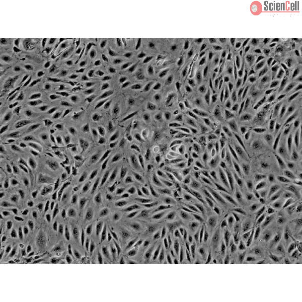
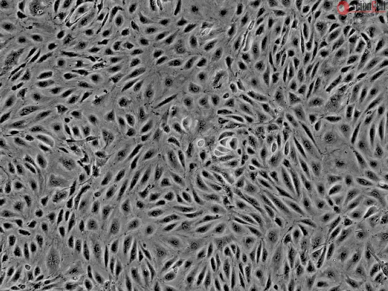
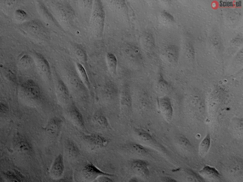
Angiogenesis is critical for many physiological processes including organ development, tissue repair, and the female reproductive tract. Human endometrial microvascular endothelial cells (HEMEC) are located in the inner mucous membrane of the uterus known as the endometrium. HEMEC are involved in endometrial angiogenesis during the menstrual cycle with the rapid growth and shedding of the endometrium. Angiogenesis in the endometrium occurs through the non-sprouting mechanisms intussusception and vessel elongation. Studies have indicated that elevated HEMEC proliferation in the endometrium can lead to disorders such as endometriosis and menorrhagia. Cultured HEMEC are a useful model to elucidate the mechanisms of normal and pathological angiogenesis and develop treatments for female reproductive tract disorders.
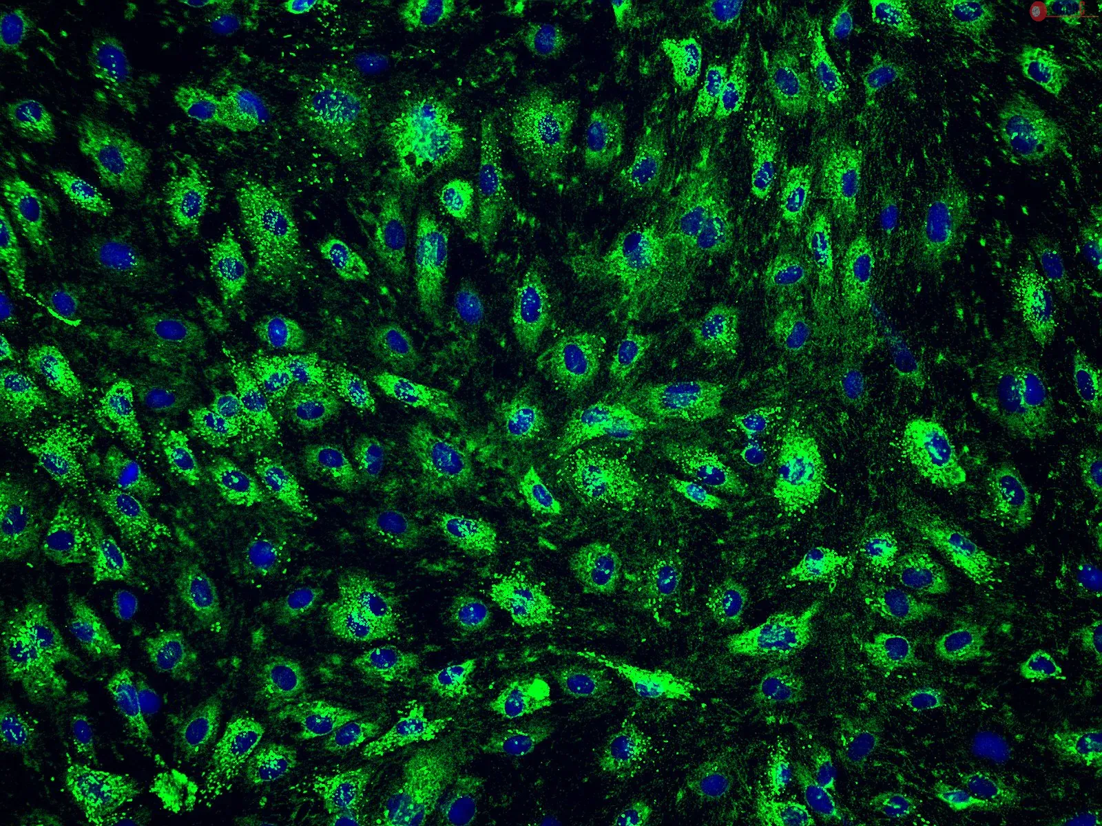
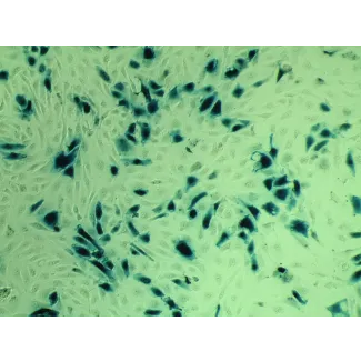
HEMEC from ScienCell Research Laboratories are isolated from human uterus. HEMEC are cryopreserved at passage one and delivered frozen. Each vial contains >5 x 10⁵ cells in 1 ml volume. HEMEC are characterized by immunofluorescence with antibodies specific to vWF/Factor VIII and/or CD31 (PECAM1). HEMEC are negative for HIV-1, HBV, HCV, mycoplasma, bacteria, yeast, and fungi. HEMEC are guaranteed to further expand for 10 population doublings under the conditions provided by ScienCell Research Laboratories.
Recommended Medium
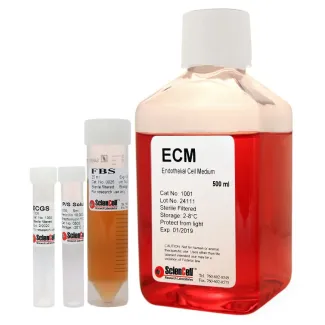
It is recommended to use Endothelial Cell Medium (ECM, Cat. #1001) for culturing HEMEC in vitro.
Product Sheets
 7010
7010
Search for a Certificate of Analysis by Lot Number below:
The endometrium undergoes rapid cycles of vascular growth, remodeling, and breakdown during the menstrual cycle and pregnancy. Decidualization is an endometrial different... More
The endometrium undergoes rapid cycles of vascular growth, remodeling, and breakdown during the menstrual cycle and pregnancy. Decidualization is an endometrial differentiation process driven by steroidal sex hormones that is critical for blastocyst-uterine interfacing and blastocyst implantation. Certain pregnancy disorders may be linked to decidualization processes. However, much remains unknown regarding the role of decidualization and reciprocal trophoblast-endometrial interactions on endometrial angiogenesis and trophoblast invasion. Here, we report an engineered endometrial microvascular network embedded in gelatin hydrogels that displays morphological and functional patterns of decidualization. Vessel complexity and biomolecule secretion are sensitive to decidualization and affect trophoblast motility, but that signaling between endometrial and trophoblast cells was not bi-directional. Although endometrial microvascular network decidualization status influences trophoblast cells, trophoblast cells did not induce structural changes in the endometrial microvascular networks. These findings add to a growing literature that the endometrium has biological agency at the uterine-trophoblast interface during implantation. Finally, we form a stratified endometrial tri-culture model, combining engineered microvascular networks with epithelial cells. These endometrial microvascular networks provide a well-characterized platform to investigate dynamic changes in angiogenesis in response to pathological and physiological endometrial states. Less
Uterine leiomyomas are the most common pelvic tumor in women of reproductive age; they cause irregular heavy menstrual bleeding leading to anemia and subsequent negative ... More
Uterine leiomyomas are the most common pelvic tumor in women of reproductive age; they cause irregular heavy menstrual bleeding leading to anemia and subsequent negative effects on quality of life. Exosomes have arisen as main players of disease progression in several illnesses, including a range of benign and malignant conditions; however, their role in leiomyomas’ pathophysiology remains unknown. We investigated the effect of exosomes derived from human uterine leiomyoma tumor cells (HULM) and human myometrial cells (UTSM) on the behavior of human endometrial microvascular endothelial cells (HEMEC). HULM- and UTSM-derived exosomes were isolated and cocultured with HEMECs. Then, cell proliferation, mRNA expression, tube formation assay, and RNA-seq were performed. Treatment of HEMEC with HULM-derived exosomes increased cell proliferation by 60% compared to control untreated cells, upregulated C-MYC and VEGFA expression levels, and increased tube formation, length, and branching (markers of angiogenesis). Profiling of miRNA revealed that 84 miRNAs were significantly downregulated and 71 were upregulated in HULM-derived exosomes compared to UTSM-derived exosomes. These findings suggest that HULM-derived exosomes might have effects on HEMEC function, containing factors that enhance endometrial proliferation and angiogenesis, which may contribute to heavy menstrual bleeding. Further research on exosomes in uterine leiomyoma may identify possible novel biomarkers for treatment. Less
ScienCell Research Laboratories (SRL) takes pride in being a resource for researchers all over the world. The publications listed here are not meant as an endorsement or confirmation of the reliability of the products.