The recombinant vesicular stomatitis virus–vectored Zaire Ebola virus glycoprotein (rVSVΔG-ZEBOV-GP) vaccine, while effective and well-tolerated, exhibits notable reac... More
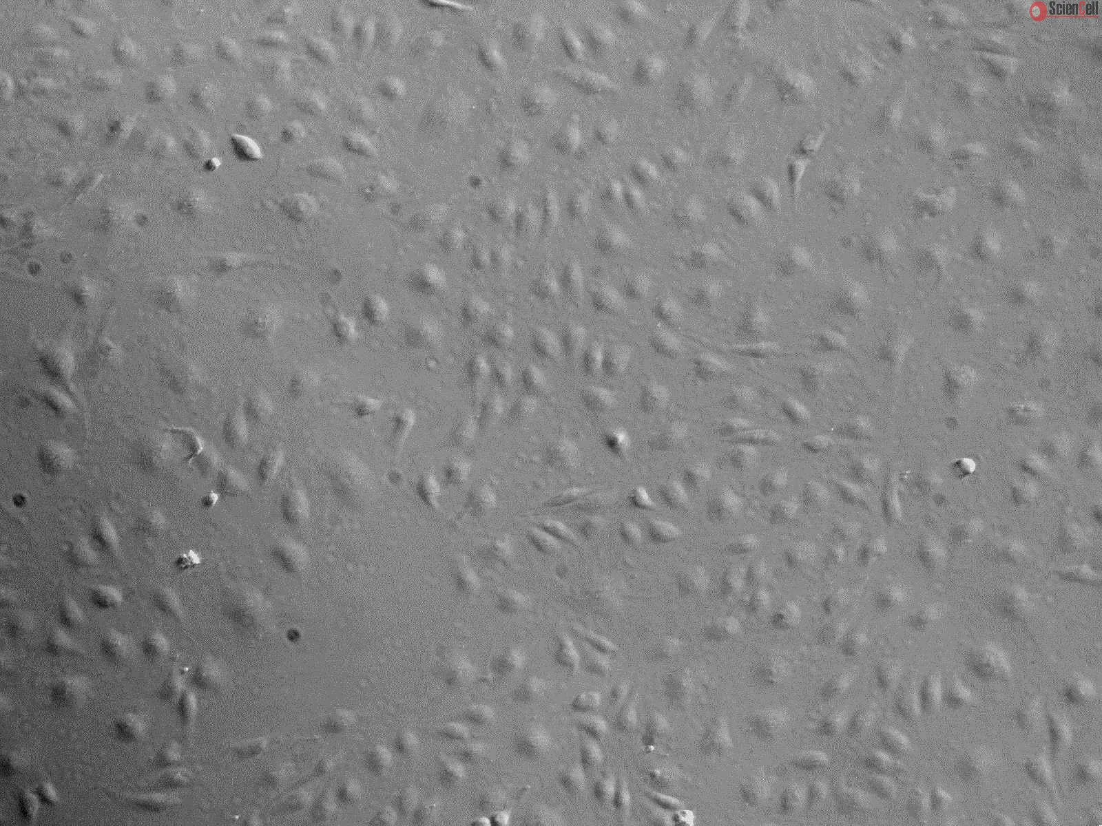
The recombinant vesicular stomatitis virus–vectored Zaire Ebola virus glycoprotein (rVSVΔG-ZEBOV-GP) vaccine, while effective and well-tolerated, exhibits notable reactogenicity, manifesting in expected adverse events (AEs), such as fever, headache, and pain, along with rare, unexpected AEs, including skin lesions, cutaneous vasculitis, and transient arthritis. The presence or absence of AEs following rVSVΔG-ZEBOV-GP vaccination is associated with a specific innate plasma signature. This study aims to elucidate in vitro the tropism of the vaccine for different cell types derived from tissues previously reported to be involved in the unexpected AEs. Upon in vitro infection with rVSVΔG-ZEBOV-GP, various cell types, such as synoviocytes, fibroblasts, keratinocytes, and endothelial cells (except chondrocytes), demonstrate productive infection, which in dermal fibroblasts triggered the release of many innate plasma signature markers, including keratinocytes’ pro-inflammatory and proapoptotic cytokines such as OSM and TRAIL. Infected monocytes from buffy coats, activated by infection, produce most innate plasma signature markers. In co-culture, rVSVΔG-ZEBOV-GP-infected monocytes serve as a source to synoviocyte infection, resulting in distinct kinetics modulation in innate biomarkers (transcription and secretion) and upregulation of specific genes, such as NEDD8 and SIGLEC-1, which have been associated with inflammatory arthritis in animal models. Altogether, our work, based on in vitro studies, provides insights into the possible mechanisms of rVSVΔG-ZEBOV-GP underlying the observed reactogenicity by showing tropism of the vaccine for off-target cells derived from AE-affected compartments (skin, joints, vessels). Furthermore, in vitro interaction with infected monocytes modulates the innate response of synoviocytes. Less
Cerebral malaria (CM) is a deadly complication of Plasmodium falciparum infection, but specific interactions involved in cerebral homing of infected erythrocytes (IEs) ar... More
Cerebral malaria (CM) is a deadly complication of Plasmodium falciparum infection, but specific interactions involved in cerebral homing of infected erythrocytes (IEs) are poorly understood. In this study, P. falciparum-IEs were characterized for binding to primary human brain microvascular endothelial cells (HBMECs). Before selection, CD36 or ICAM-1-binding parasites exhibited punctate binding to a subpopulation of HBMECs and binding was CD36 dependent. Panning of IEs on HBMECs led to a more dispersed binding phenotype and the selection of three var genes, including two that encode the tandem domain cassette 8 (DC8) and were non-CD36 binders. Multiple domains in the DC8 cassette bound to brain endothelium and the cysteine-rich interdomain region 1 inhibited binding of P. falciparum-IEs by 50%, highlighting a key role for the DC8 cassette in cerebral binding. It is mysterious how deadly binding variants are maintained in the parasite population. Clonal parasite lines expressing the two brain-adherent DC8-var genes did not bind to any of the known microvascular receptors, indicating unique receptors are involved in cerebral binding. They could also adhere to brain, lung, dermis, and heart endothelial cells, suggesting cerebral binding variants may have alternative sequestration sites. Furthermore, young African children with CM or nonsevere control cases had antibodies to HBMEC-selected parasites, indicating they had been exposed to related variants during childhood infections. This analysis shows that specific P. falciparum erythrocyte membrane protein 1 types are linked to cerebral binding and suggests a potential mechanism by which individuals may build up immunity to severe disease, in the absence of CM. Less
Cerebral malaria is the most deadly manifestation of infection with Plasmodium falciparum. The pathology of cerebral malaria is characterized by the accumulation of infec... More
Cerebral malaria is the most deadly manifestation of infection with Plasmodium falciparum. The pathology of cerebral malaria is characterized by the accumulation of infected erythrocytes (IEs) in the microvasculature of the brain caused by parasite adhesins on the surface of IEs binding to human receptors on microvascular endothelial cells. The parasite and host molecules involved in this interaction are unknown. We selected three P. falciparum strains (HB3, 3D7, and IT/FCR3) for binding to a human brain endothelial cell line (HBEC-5i). The whole transcriptome of isogenic pairs of selected and unselected parasites was analyzed using a variant surface antigen-supplemented microarray chip. After selection, the most highly and consistently up-regulated genes were a subset of group A-like var genes (HB3var3, 3D7_PFD0020c, ITvar7, and ITvar19) that showed 11- to >100-fold increased transcription levels. These var genes encode P. falciparum erythrocyte membrane protein (PfEMP)1 variants with distinct N-terminal domain types (domain cassette 8 or domain cassette 13). Antibodies to HB3var3 and PFD0020c recognized the surface of live IEs and blocked binding to HBEC-5i, thereby confirming the adhesive function of these variants. The clinical in vivo relevance of the HBEC-selected parasites was supported by significantly higher surface recognition of HBEC-selected parasites compared with unselected parasites by antibodies from young African children suffering cerebral malaria (Mann-Whitney test, P = 0.029) but not by antibodies from controls with uncomplicated malaria (Mann-Whitney test, P = 0.58). This work describes a binding phenotype for virulence-associated group A P. falciparum erythrocyte membrane protein 1 variants and identifies targets for interventions to treat or prevent cerebral malaria. Less
Objectives: Angiogenesis is closely associated with osteogenesis where reciprocal interactions between endothelial and osteoblast cells play an important role in bone reg... More
Objectives: Angiogenesis is closely associated with osteogenesis where reciprocal interactions between endothelial and osteoblast cells play an important role in bone regeneration. For these reasons, the aim of this work was to develop a co-culture system to study in detail any time-dependent interactions between human mesenchymal stem cells (HMSC) and human dermal microvascular endothelial cells (HDMEC), co-cultured in a 2D system, for 35 days. Materials and methods: HMSC and HDMEC were co-cultured at a ratio of 1:4, respectively. Single-cell cultures were used as controls. Cell viability/proliferation was assessed using MTT, DNA quantification and calcein-AM assays. Cell morphology was monitored using confocal microscopy, and real time PCR was performed. Alkaline phosphatase activity and histochemical staining were evaluated. Matrix mineralization assays were also performed. Results: Cells were able to grow in characteristic patterns maintaining their viability and phenotype expression throughout culture time, compared to HMSC and HDMEC monocultures. HMSC differentiation seemed to be enhanced in the co-culture conditions, since it was observed an over expression of osteogenesis-related genes, and of ALP activity. Furthermore, presence of calcium phosphate deposits was also confirmed. Conclusions: This work reports in detail the interactions between HMSC and HDMEC in a long-term co-culture 2D system. Endothelial and mesenchymal stem cells cultured in the present co-culture conditions ensured proliferation and phenotype differentiation of cell types, osteogenesis stimulation and over-expression of angiogenesis-related genes, in the same culture system. It is believed that the present work can lead to significant developments for bone tissue regeneration and cell biology studies. © 2012 Blackwell Publishing Ltd. Less
The chromosomal region 10p13 has been linked to paucibacillary leprosy in two independent studies. The MRC1 gene, encoding the human mannose receptor (MR), is located in ... More
The chromosomal region 10p13 has been linked to paucibacillary leprosy in two independent studies. The MRC1 gene, encoding the human mannose receptor (MR), is located in the 10p13 region and non-synonymous SNPs in exon 7 of the gene have been suggested as leprosy susceptibility factors. We determined that G396S is the only non-synonymous exon 7-encoded polymorphism in 396 unrelated Vietnamese subjects. This SNP was genotyped in 490 simplex and 90 multiplex leprosy families comprising 704 patients (47% paucibacillary; 53% multibacillary). We observed significant under-transmission of the serine allele of the G396S polymorphism with leprosy per se (P = 0.036) and multibacillary leprosy (P = 0.034). In a sample of 384 Brazilian leprosy cases (51% paucibacillary; 49% multibacillary) and 399 healthy controls, we observed significant association of the glycine allele of the G396S polymorphism with leprosy per se (P = 0.016) and multibacillary leprosy (P = 0.023). In addition, we observed a significant association of exon 7 encoded amino acid haplotypes with leprosy per se (P = 0.012) and multibacillary leprosy (P = 0.004). Next, we tested HEK293 cells over-expressing MR constructs (293-MR) with three exon 7 haplotypes of MRC1 for their ability to bind and internalize ovalbumin and zymosan, two classical MR ligands. No difference in uptake was measured between the variants. In addition, 293-MR failed to bind and internalize viable Mycobacterium leprae and BCG. We propose that the MR-M. leprae interaction is modulated by an accessory host molecule of unknown identity. Less
Laser-induced vessel wall injury leads to rapid thrombus formation in an animal thrombosis model. The target of laser injury is the endothelium. We monitored calcium mobi... More
Laser-induced vessel wall injury leads to rapid thrombus formation in an animal thrombosis model. The target of laser injury is the endothelium. We monitored calcium mobilization to assess activation of the laser-targeted cells. Infusion of Fluo-4 AM, a calcium-sensitive fluorochrome, into the mouse circulation resulted in dye uptake in the endothelium and circulating hematopoietic cells. Laser injury in mice treated with eptifibatide to inhibit platelet accumulation resulted in rapid calcium mobilization within the endothelium. Calcium mobilization correlated with the secretion of lysosomal-associated membrane protein 1, a marker of endothelium activation. In the absence of eptifibatide, endothelium activation preceded platelet accumulation. Laser activation of human umbilical vein endothelial cells loaded with Fluo-4 resulted in a rapid increase in calcium mobilization associated cell fluorescence similar to that induced by adenosine diphosphate (10 μM) or thrombin (1 U/mL). Laser activation of human umbilical vein endothelial cells in the presence of corn trypsin inhibitor treated human plasma devoid of platelets and cell microparticles led to fibrin formation that was inhibited by an inhibitory monoclonal anti-tissue factor antibody. Thus laser injury leads to rapid endothelial cell activation. The laser activated endothelial cells can support formation of tenase and prothrombinase and may be a source of activated tissue factor as well. Less
Oncolytic viral (OV) therapy is a promising therapeutic modality for brain tumors. Vasculostatin (Vstat120) is the cleaved and secreted extracellular fragment of brain-sp... More
Oncolytic viral (OV) therapy is a promising therapeutic modality for brain tumors. Vasculostatin (Vstat120) is the cleaved and secreted extracellular fragment of brain-specific angiogenesis inhibitor 1 (BAI1), a brain-specific receptor. To date, the therapeutic efficacy of Vstat120 delivery into established tumors has not been investigated. Here we tested the therapeutic efficacy of combining Vstat120 gene delivery in conjunction with OV therapy. We constructed RAMBO (Rapid Antiangiogenesis Mediated By Oncolytic virus), which expresses Vstat120 under the control of the herpes simplex virus (HSV) IE4/5 promoter. Secreted Vstat120 was detected as soon as 4 hours postinfection in vitro and was retained for up to 13 days after OV therapy in subcutaneous tumors. RAMBO-produced Vstat120 efficiently inhibited endothelial cell migration and tube formation in vitro (P = 0.0005 and P = 0.0184, respectively) and inhibited angiogenesis (P = 0.007) in vivo. There was a significant suppression of intracranial and subcutaneous glioma growth in mice treated with RAMBO compared to the control virus, HSVQ (P = 0.0021 and P < 0.05, respectively). Statistically significant reduction in tumor vascular volume fraction (VVF) and microvessel density (MVD) was observed in tumors treated with RAMBO. This is the first study to report the antitumor effects of Vstat120 delivery into established tumors and supports the further development of RAMBO as a possible cancer therapy. Less
Although the liver is known to be the main site of factor VIII (FVIII) production, other organs are probably also important for the regulation of FVIII secretion. However... More
Although the liver is known to be the main site of factor VIII (FVIII) production, other organs are probably also important for the regulation of FVIII secretion. However, the study of the regulation of extrahepatic FVIII production has been hampered by the lack of definitive identification of human tissues able to secrete FVIII. Recent studies have shown that lung endothelial cells can synthesize FVIII. We therefore studied the production of FVIII by endothelial cells purified from other vascular beds. Because physiologic stress results in a rapid elevation of FVIII, we also investigated whether endothelial cells can store FVIII and secrete it after treatment with agonists. Microvascular endothelial cells from lung, heart, intestine, and skin as well as endothelial cells from pulmonary artery constitutively secreted FVIII and released it after treatment with phorbol-myristate acetate and epinephrine. By contrast, endothelial cells from the aorta, umbilical artery and umbilical vein did not constitutively secrete FVIII or release it after treatment with agonists, probably because of a lack of FVIII synthesis. Extrahepatic endothelial cells from certain vascular beds therefore appear to be an important FVIII production and storage site with the potential to regulate FVIII secretion in chronic and acute conditions. Less
Heat shock protein 90alpha (Hsp90alpha) is a ubiquitously expressed molecular chaperone, which is essential for the maintenance of eukaryote homeostasis. Hsp90alpha can a... More
Heat shock protein 90alpha (Hsp90alpha) is a ubiquitously expressed molecular chaperone, which is essential for the maintenance of eukaryote homeostasis. Hsp90alpha can also be secreted extracellularly and is associated with several physiological and pathological processes including wound healing, cancer, infectious diseases and diabetes. Angiogenesis, defined as the sprouting of new blood vessels from pre-existing capillaries via endothelial cell proliferation and migration, commonly occurs in and contributes to the above mentioned processes. However, the secretion of Hsp90alpha from endothelial cells and also its function in angiogenesis are still unclear. Here we investigated the role of extracellular Hsp90alpha in angiogenesis using dermal endothelial cells in vitro and a wound healing model in vivo. We find that the secretion of Hsp90alpha but not Hsp90beta is increased in activated endothelial cells with the induction of angiogenic factors and matrix proteins. Secreted Hsp90alpha localizes on the leading edge of endothelial cells and promotes their angiogenic activities, whereas Hsp90alpha neutralizing antibodies reverse the effect. Furthermore, using a mouse skin wound healing model in vivo, we demonstrate that extracellular Hsp90alpha localizes on blood vessels in granulation tissues of wounded skin and promotes angiogenesis during wound healing. Taken together, our study reveals that Hsp90alpha can be secreted by activated endothelial cells and is a positive regulator of angiogenesis, suggesting the potential application of Hsp90alpha as a stimulator for wound repair. Copyright 2010 Elsevier Inc. All rights reserved. Less
Background: Alveolar cell senescence is accelerated in patients with chronic obstructive pulmonary disease (COPD). Objectives: We tested the hypothesis that alveolar cell... More
Background: Alveolar cell senescence is accelerated in patients with chronic obstructive pulmonary disease (COPD). Objectives: We tested the hypothesis that alveolar cell senescence contributes to the chronic inflammation that affects the lungs of COPD patients. Methods: We exposed alveolar type II-like epithelial (A549) cells to a G-quadruplex-interacting telomerase inhibitor in vitro to induce cellular senescence and analyzed the production of proinflammatory cytokines and the activation of NF-kappaB. Human dermal microvascular endothelial cells (HDMECs) were serially passaged to induce replicative senescence. We also immunostained human lung tissue sections obtained from COPD patients, asymptomatic smokers and asymptomatic nonsmokers and examined correlations between type II cell senescence and inflammation. Results: Senescent A549 cells and HDMECs, whether stimulated with lipopolysaccharide or not, produced greater amounts of IL-6, IL-8 and TNF-alpha, which paralleled NF-kappaB activation, than did presenescent cells. There were positive correlations between the percentages of senescent type II cells that expressed p16(INK4a) and the percentages of type II cells that expressed phosphorylated NF-kappaB. The lung tissue of the COPD patients contained higher percentages of proinflammatory senescent type II cells that co-expressed p16(INK4a) and phosphorylated NF-kappaB than the tissue from asymptomatic smokers and asymptomatic nonsmokers. Higher percentages of p16(INK4a)-positive senescent type II cells than of p16(INK4a)-negative presenescent type II cells were positive for phosphorylated NF-kappaB. Conclusions: Senescence of alveolar epithelial cells is associated with functional alterations of the cells to a proinflammatory phenotype and may contribute to the pathogenesis of COPD. Less
To determine how ascorbic acid moves from the bloodstream into tissues, we assessed transfer of the vitamin across the barrier generated by EA.hy926 endothelial cells whe... More
To determine how ascorbic acid moves from the bloodstream into tissues, we assessed transfer of the vitamin across the barrier generated by EA.hy926 endothelial cells when these were cultured on semipermeable filter supports. Ascorbate transfer from the luminal to the abluminal compartment was time dependent, inhibited by anion channel blockers and by activation of protein kinase A, but was increased by thrombin. Ascorbate transfer occurred by a paracellular route, since it did not correlate with intracellular ascorbate contents and was not rectified or saturable. Nonetheless, intracellular ascorbate inhibited the transfer of both ascorbate and radiolabeled inulin across the endothelial barrier. The increase in barrier function due to ascorbate was dependent on its intracellular concentration, significant by 15 min of incubation, prevented by the cytoskeletal inhibitor colchicine, associated with F-actin stress fiber formation, and not due to collagen deposition. These results show that ascorbate traverses the endothelial barrier by a paracellular route that is regulated by cell metabolism, ion channels, and ascorbate itself. Since the latter effect occurred over the physiological range of ascorbate plasma concentrations, it could reflect a role for the vitamin in control of endothelial barrier function in vivo. Keywords: paracellular transport, ascorbate transport, anion channels, endothelial permeability Less
Nitrite (NO(2)(-)) recycling to nitric oxide (NO) is catalysed by a number of enzymes and induces a protective vasodilation effect under hypoxia/ischaemia. In the present... More
Nitrite (NO(2)(-)) recycling to nitric oxide (NO) is catalysed by a number of enzymes and induces a protective vasodilation effect under hypoxia/ischaemia. In the present work, we tested the in vitro ability of the three NOS (nitric oxide synthase) isoforms to release NO from nitrite under anoxia using electrochemical detection, chemiluminescence and absorption spectroscopy. The release of free NO from anoxic nitrite solutions at 15 muM was specific to the endothelial NOS isoform (eNOS) and did not occur with the neuronal (nNOS) or inducible (iNOS) isoforms. Unlike xanthine oxidase, the eNOS reductase domain did not recycle nitrite to NO, and wild-type eNOS did not reduce nitrate. Our data suggest that structural and, by inference, dynamic differences between nNOS and eNOS in the distal haem side account for eNOS being the only isoform capable of converting nitrite into NO at pH 7.6. In human dermal microvascular endothelial cells under careful control of oxygen tension, the rates of NO formation determined by chemiluminescence were enhanced approximately 3.6- and approximately 8.3-fold under hypoxia (2 p.p.m. O(2)) and anoxia (argon) respectively compared with normoxia ( approximately 22 p.p.m. O(2)) using 10 muM extracellular nitrite. NOS inhibitors inhibited this hypoxic NO release. Our data show that eNOS is unique in that it releases NO under all oxygen levels from normoxia to complete anoxia at physiological micromolar nitrite concentrations. The magnitude of the hypoxic NO release by the endothelial cells suggest that the endothelium could provide an appropriate response to acute episodic ischaemia and may explain the observed eNOS-expression-specific protective effect as a short-term response in animal models of acute hypoxia. Less
Pericytes play a crucial role in the homeostasis and maturation of newly formed vessels, but their precursor and the process of their differentiation remain unclear. In t... More
Pericytes play a crucial role in the homeostasis and maturation of newly formed vessels, but their precursor and the process of their differentiation remain unclear. In this study, we show that in vivo, pericytes in human granulation tissue taken from burn patients expressed CD13 and collagen I, which are cell markers of peripheral blood fibrocytes (PBFCs). Mouse PBFCs, ex vivo labeled by PKH-26 and administered intravenously back to mice, formed lumens in the granulation tissue and expressed pericyte marker NG2. Furthermore, in cell culture, human PBFCs in co-culture with human umbilical vascular endothelial cells or human dermal microvascular endothelial cells (HDMECs) expressed pericyte markers desmin and myosin heavy chain of smooth muscle cell, and some migrated to the membrane of human umbilical vascular endothelial cells or HDMECs to form PBFC/vascular endothelial cell clusters, or formed round PBFC only clusters with their nuclei aligned along a circle. The formation of PBFC/vascular endothelial cell clusters and round PBFC clusters were inhibited by an antibody against monocyte chemotactic protein-1. PBFCs cultured with homogenate of granulation tissue also expressed desmin and myosin heavy chain of smooth muscle cell. Our findings support that the PBFC is a precursor of pericytes, the differentiation of PBFCs is induced by vascular endothelial cells in a paracrine fashion. Less


.webp)