Lymphangiogenesis in inflammation has received considerable attention in recent years. Administration of modulating lymphangiogenesis provides more possibilities of treat... More
Lymphangiogenesis in inflammation has received considerable attention in recent years. Administration of modulating lymphangiogenesis provides more possibilities of treating inflammation-associated diseases. However, the main mediators and factors governing inflammation-induced lymphangiogenesis (ILA) are yet to be defined. Here, we explored the role of HMGB1-TLR4 signalling pathway in modulating inflammation-induced lymphangiogenesis and its underlying mechanisms using an ILA mouse model and 2 cell lines. Our results show that HMGB1 promoted VEGF-C-induced HDLECs proliferation in a dose-dependent manner and TLR4 mediates HMGB1-induced LECs proliferation and tube formation in vitro. And in vivo, rHMGB1 treatment significantly promoted ILA, and the promoting effects was inhibited notably when HMGB1-TLR4 was blocked. HMGB1-associated ILA is primarily dependent on TLR4 but not on TLR2. In mechanisms, the recruitment and activation of CD11b+ cells are important cellular mechanisms in HMGB1-TLR4 associated ILA, and multiple key pro-lymphangiogenesis molecules mediates HMGB1-TLR4 associated ILA, including VEGF-C/VEGFR3, inflammatory factors IL-1β and TNF-α, MMP-2 and MMP-9 and NF-κB p65. In conclusion, HMGB1-associated ILA is primarily dependent on TLR4, and CD11b+ cells and multiple molecular mechanisms mediate HMGB1-TLR4 associated ILA. Furthermore, the ILA can be effectively modulated by HMGB1-TLR4 signalling. Consequently, administration of modulating ILA through HMGB1-TLR4 pathway may provide us more possibilities of treating inflammation and lymphangiogenesis associated diseases. Less
Background CC-chemokine receptor 7 (CCR7), which plays an important role in cell directional movement, is highly expressed in various cancers and positively related to ly... More
Background CC-chemokine receptor 7 (CCR7), which plays an important role in cell directional movement, is highly expressed in various cancers and positively related to lymph node metastasis. The inflammatory cytokine tumour necrosis factor (TNF)-α promotes tumour progression and lymph node metastasis in gallbladder cancer (GBC). However, the expression of CCR7 in GBC is unclear, and its role in the TNF-α-induced lymphatic metastasis of GBC requires further research. Methods The expression of CCR7 in clinical samples was detected by immunohistochemistry, and the relationship between CCR7 and clinicopathological factors or the TNF-α level of the bile was analyzed. After treatment with various concentrations of TNF-α, CCR7 expression in GBC cell lines was measured by Western blotting. The relative luciferase reporter assay, site-directed mutagenesis and chromatin immunoprecipitation were used to analyze the promoter activity and transcriptional regulation of CCR7. MAPKs inhibitors were used to explore the upstream signalling molecules of AP-1. We established a NOZ cell line stably expressing lentiviral CCR7 shRNA that effectively silenced the expression of CCR7, and to determine the role of TNF-α - CCR7 axis in the migration of GBC cells to the lymphatic system by transwell assays and animal experiments. Results CCR7 was highly expressed in GBC samples. Higher expression of CCR7 was associated with American Joint Committee on Cancer (AJCC) staging and lymph node metastasis. Moreover, we found that CCR7 expression in GBC tissue was positively correlated with the levels of TNF-α in the bile, and that TNF-α enhanced the promoter activity and protein expression of CCR7 through the “ERK1/2-AP-1” and “JNK-AP-1” pathways. Finally, we revealed that TNF-α could promote GBC cell migration to lymphatic endothelial cells or lymph nodes through upregulation of CCR7 in vitro and in vivo. Conclusions Our study suggests that CCR7 is highly expressed in GBC, and mediates the TNF-α-induced lymphatic metastasis of GBC through the “TNF-α - ERK1/2 - AP-1 - CCR7” and “TNF-α - JNK - AP-1 - CCR7” pathways. Less
Background Tumor necrosis factor-alpha (TNF-α), a key player in cancer-related inflammation, was recently demonstrated to be involved in the lymphatic metastasis of gall... More
Background Tumor necrosis factor-alpha (TNF-α), a key player in cancer-related inflammation, was recently demonstrated to be involved in the lymphatic metastasis of gallbladder cancer (GBC). Vascular endothelial growth factor D (VEGF-D) is a key lymphangiogenic factor that is associated with lymphangiogenesis and lymph node metastasis in GBC. However, whether VEGF-D is involved in TNF-α-induced lymphatic metastasis of GBC remains undetermined. Methods The expression of VEGF-D in patient specimens was detected by immunohistochemistry and the relationship between VEGF-D in the tissue and TNF-α in the bile of the matching patients was analyzed. The VEGF-D mRNA and protein levels after treatment with exogenous TNF-α in NOZ, GBC-SD and SGC-996 cell lines were measured by real-time PCR and ELISA. The promoter activity and transcriptional regulation of VEGF-D were analyzed with the relative luciferase reporter assay, mutant constructs, electrophoretic mobility shift assay (EMSA), chromatin immunoprecipitation (ChIP) assay, RNA interference and Western blotting. Inhibitors of JNK, p38 MAPK and ERK1/2 were used to explore the upstream signaling effector of AP-1. We used lentiviral vector expressing a VEGF-D shRNA construct to knockdown VEGF-D gene in NOZ and GBC-SD cells. The role of the TNF-α-VEGF-D axis in the tube formation of human dermal lymphatic endothelial cells (HDLECs) was determined using a three-dimensional coculture system. The role of the TNF-α - VEGF-D axis in lymphangiogenesis and lymph node metastasis was studied via animal experiment. Results TNF-α levels in the bile of GBC patients were positively correlated with VEGF-D expression in the clinical specimens. TNF-α can upregulate the protein expression and promoter activity of VEGF-D through the ERK1/2 - AP-1 pathway. Moreover, TNF-α can promote tube formation of HDLECs, lymphangiogenesis and lymph node metastasis of GBC by upregulation of VEGF-D in vitro and in vivo. Conclusion Taken together, our data suggest that TNF-α can promote lymphangiogenesis and lymphatic metastasis of GBC through the ERK1/2/AP-1/VEGF-D pathway. Keyword: Gallbladder cancer, TNF-α, VEGF-D, Lymphatic metastasis Less
In contrast to blood capillaries, lymphatic capillaries in peripheral tissues are composed of a single-cell layer of lymphatic endothelial cells (LECs) without a covering... More
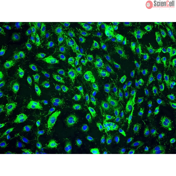
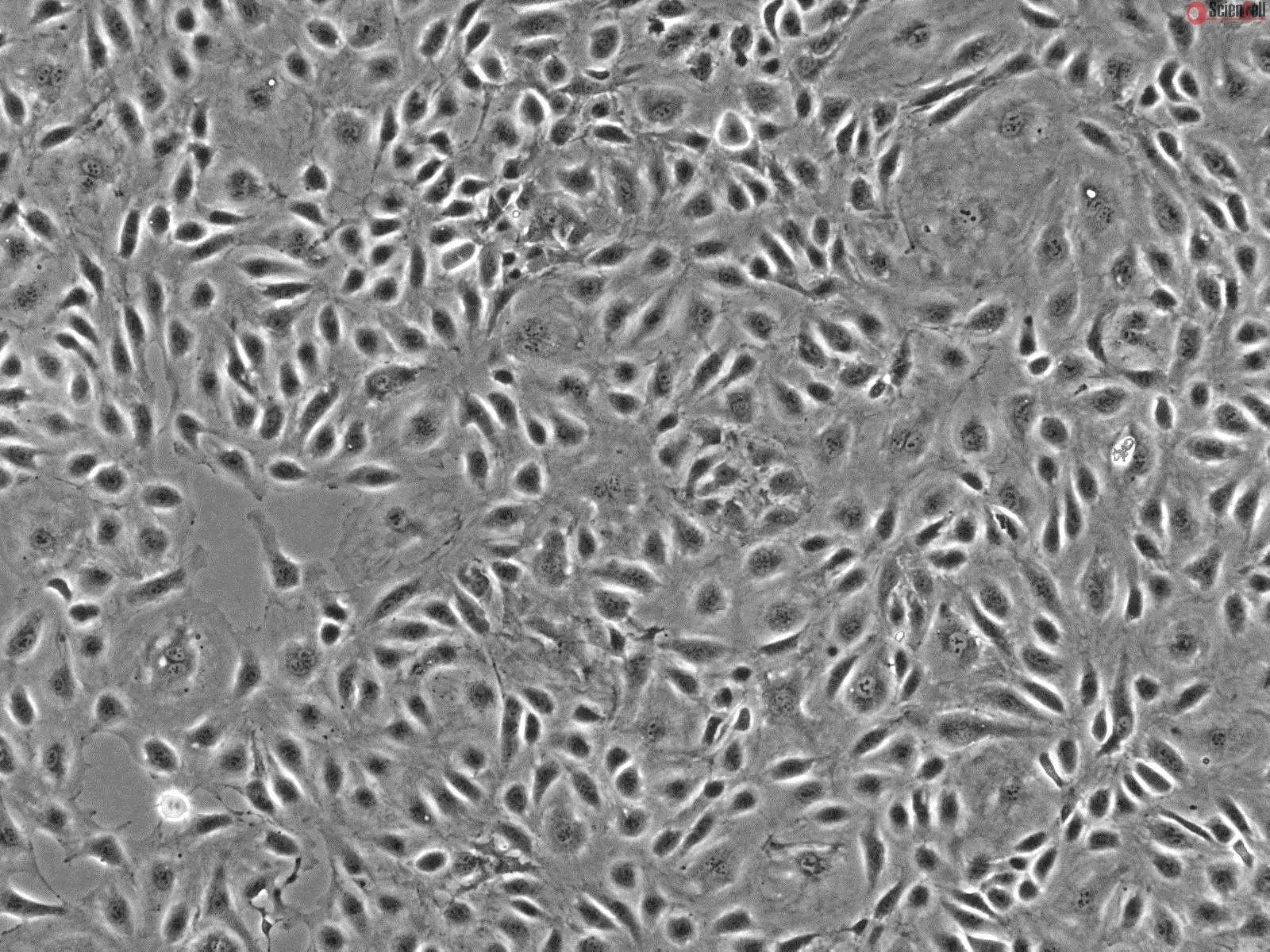
In contrast to blood capillaries, lymphatic capillaries in peripheral tissues are composed of a single-cell layer of lymphatic endothelial cells (LECs) without a covering of mural cells. However, in lymphatic malformations, the enlarged lymphatic vessels were covered with mural cells. This study aimed to understand the molecular mechanism of differences between human dermal lymphatic endothelial cells (HDLECs) and human umbilical vein endothelial cells (HUVECs) and to determine the changes of LECs in the pathological condition of lymphatic malformation. Results showed that HDLECs exhibited lower expression of endothelial proteins, including VE-cadherin and CD31, than HUVECs; HDLECs also showed higher expression of mesenchymal proteins, including α-SMA, SM22α, calponin, and epithelial mesenchymal transition-related transcription factor Slug, than HUVECs. Likewise, HDLECs displayed higher permeability and weaker recruitment of SMCs than HUVECs; HDLECs also exhibited low PDGF-BB expression. TGF-β2 treatment and FGF2 depletion enhanced mesenchymal marker expression with increased permeability and reduced SMC recruitment. By contrast, Slug depletion in HDLECs enhanced VE-cadherin expression, inhibited α-SMA expression, decreased permeability, and enhanced PDGF-BB expression. These results suggested that HDLECs were in a mesenchymal status, which contributed to their functions and might determine their identities. Our data also revealed that miR143/145 was implicated in the mesenchymal status of HDLECs. In lymphatic malformations (LMs) treated with OK-432 sclerotherapy, immunohistochemistry results showed that Prox1 expression was reduced and mural cell investment was increased; these results indicated that LECs lost their mesenchymal status after OK-432 treatment was administered. The decreased mesenchymal status of LECs in LMs may induce dilated vessel constriction, which could be the mechanism of OK-432 sclerotherapy Less
Autophagy, a type II programmed cell death, is essential for cell survival under stress, e.g. lung injury, and bone marrow-derived mesenchymal stem cells (BM-MSCs) have g... More
Autophagy, a type II programmed cell death, is essential for cell survival under stress, e.g. lung injury, and bone marrow-derived mesenchymal stem cells (BM-MSCs) have great potential for cell therapy. However, the mechanisms underlying the BM-MSC activation of autophagy to provide a therapeutic effect in ischaemia/reperfusion-induced lung injury (IRI) remain unclear. Thus, we investigate the activation of autophagy in IRI following transplantation with BM-MSCs. Seventy mice were pre-treated with BM-MSCs before they underwent lung IRI surgery in vivo. Human pulmonary micro-vascular endothelial cells (HPMVECs) were pre-conditioned with BM-MSCs by oxygen-glucose deprivation/reoxygenation (OGD) in vitro. Expression markers for autophagy and the phosphoinositide 3-kinase/protein kinase B (PI3K/Akt) signalling pathway were analysed. In IRI-treated mice, administration of BM-MSCs significantly attenuated lung injury and inflammation, and increased the level of autophagy. In OGD-treated HPMVECs, co-culture with BM-MSCs attenuated endothelial permeability by decreasing the level of cell death and enhanced autophagic activation. Moreover, administration of BM-MSCs decreased the level of PI3K class I and p-Akt while the expression of PI3K class III was increased. Finally, BM-MSCs-induced autophagic activity was prevented using the inhibitor LY294002. Administration of BM-MSCs attenuated lung injury by improving the autophagy level via the PI3K/Akt signalling pathway. These findings provide further understanding of the mechanisms related to BM-MSCs and will help to develop new cell-based therapeutic strategies in lung injury. Less
Background Tumor lymphangiogenesis plays an important role in promoting growth and metastasis of tumors, but no antilymphangiogenic agent is used clinically. Based on the... More
Background Tumor lymphangiogenesis plays an important role in promoting growth and metastasis of tumors, but no antilymphangiogenic agent is used clinically. Based on the effect of norcantharidin (NCTD) on lymphangiogenesis of human lymphatic endothelial cells (LECs), we firstly investigated the antilymphangiogenic activity of NCTD as a tumor lymphangiogenic inhibitor for human colonic adenocarcinomas (HCACs). Methods In vivo and in vitro experiments to determine the effects of NCTD on tumor growth and lymphangiogenesis of the in-situ colonic xenografts in nude mice, and lymphatic tube formation of the three-dimensional (3-D) of the co-culture system of HCAC HT-29 cells and LECs were done. Proliferation, apoptosis, migration, invasion, Ki-67, Bcl-2 and cell cycle of LECs and the co-culture system in vitro were respectively determined. Streparidin-peroxidase staining, SABC, western blotting and RT-PCR were respectively used to examine the expression of LYVE-1, D2-40, CK20 (including their LMVD), and VEGF-A, VEGF-C, VEGF-D, VEGFR-2 and VEGFR-3 in vitro and in vivo. Results NCTD inhibited tumor growth and lymphangiogenesis of the in-situ colonic xenografts in vivo, and these observations were confirmed by facts that lymphatic tube formation, proliferation, apoptosis, migration, invasion, S-phase cell cycle, and Ki-67 and Bcl-2 expression in vitro, and LYVE-1, D2-40, CK20 expression and their LMVD in vitro and in vivo were inhibited and affected. Furthermore, the expression of VEGF-A, VEGF-C, VEGF-D, VEGFR-2 and VEGFR-3 at protein/mRNA levels in the process of lymphatic tube formation in vitro and tumor lymphangiogenesis in vivo was downregulated; NCTD in combination with mF4-31C1 or Sorafenib enhanced these effects. Conclusions NCTD inhibits tumor growth and lymphangiogenesis of HCACs through “multi-points priming” mechanisms i.e. affecting related malignant phenotypes, inhibiting Ki-67 and Bcl-2 expression, inducing S-phase cell cycle arrest, and directly or indirectly downregulating VEGF-A,-C,-D/VEGFR-2,-3 signaling pathways. The present finding strongly suggests that NCTD could serve as a potential antilymphangiogenic agent for tumor lymphangiogenesis and is of importance to explore NCTD is used for antitumor metastatic comprehensive therapy for HCACs. Keywords: Colonic neoplasm, Norcantharidin, Tumor growth, Lymphangiogenesis, Antilymphangiogenic therapy Less
Axillary dissection during breast cancer surgery produces extensive lymphatic vessel damage that often leads to life-long secondary lymphedema of the arm. We have develop... More
Axillary dissection during breast cancer surgery produces extensive lymphatic vessel damage that often leads to life-long secondary lymphedema of the arm. We have developed a biodegradable material conduit for lymphatic vessel reconstruction where fibers electrospun along the conduit lumen promote endothelial cell alignment and migration, in vitro. The diameter and density of the electrospun fibers were optimized for cell migration and direction on 2-dimensional substrates by seeding human lymphatic endothelial cells (LECs) onto aligned fibers of varying diameters and densities, randomly oriented fibers, and film substrates with no fibers. We found that LECs became aligned in the fiber direction, with cells seeded on the randomly oriented fibers becoming oriented in random directions, whereas cells seeded on the highly aligned fibers became highly aligned. Cell migration was dependent upon fiber alignment and density, with optimal migration found on 1300 nm diameter aligned fibers of low density. Blood endothelial cells seeded on the fibers exhibited similar behavior as the LECs. Fiber alignment was preserved upon rolling the 2-dimensional substrate into the tubular geometry of a lymphatic vessel. The data suggest that aligned electrospun fibers may promote endothelial migration across the conduit in a manner that is independent of lymphatic growth factors. Less
Tumor‐associated lymphatic endothelial cells (TLEC) could play a key role in the process of tumor metastasis. The aim of this study was to investigate the effect of TLE... More
Tumor‐associated lymphatic endothelial cells (TLEC) could play a key role in the process of tumor metastasis. The aim of this study was to investigate the effect of TLECs that were isolated from human epithelial ovarian tumor (EOT) on ovarian cancer cell line CAOV‐3 in vitro. First, TLECs in EOT were detected by immunochemistry and flow cytometry, then marked by lymphatic endothelial cell (LEC) marker LYVE‐1, isolated by magnetic beads, and cultured in vitro. The cells were identified by immunostaining of LEC markers LYVE‐1, Prox‐1, Podoplanin, VEGFR‐3, and pan‐endothelial cell marker CD31. TLECs from EOT can be detected, cultured, and identified in vitro successfully. The effects of TLECs on invasion and migration of CAOV‐3 cells were investigated by 12‐well Boyden chamber; the proliferation effect was studied by counting the Trypan blue exclusion cell number. Furthermore, changes in MMP‐2/9 secreted by CAOV‐3 cells treated with TLEC were shown using real‐time PCR and zymography, and TIMP‐1/2 was detected by real‐time PCR. In vitro, TLECs can enhance invasion and migration of CAOV‐3 cells, but have no significant effect on proliferation. It was clear that the expression of MMP‐9 increased and TIMP‐2 decreased in CAOV‐3 cells treated by TLECs, and the increasing of MMP‐9 was confirmed by zymography. TLECs from EOT can enhance migration and invasion of human ovarian carcinoma cell line in vitro, and the possible mechanism was through activation of MMP‐9/TIMP‐2. Less
The major cause of cancer mortality is the metastatic spread of tumor cells that can occur via multiple routes, including the vascular system and the lymphatic system. In... More
The major cause of cancer mortality is the metastatic spread of tumor cells that can occur via multiple routes, including the vascular system and the lymphatic system. In this study, we developed an IgG-like fusion protein molecule [vascular endothelial growth factor (VEGF) receptor 31-immunoglobulin (VEGFR31-Ig)] which could simultaneously bind the angiogenic growth factor VEGF-A and the lymphangiogenic growth factor VEGF-C. Importantly, VEGFR31-Ig exhibited VEGF-A-binding affinity similar to that of VEGFTrap, the most potent VEGF-A binder, and VEGF-C-binding affinity comparable with that of the soluble fusion protein VEGFR3-Ig (sVEGFR3). Pharmacokinetic analysis in mice showed that VEGFR31-Ig had improved pharmacokinetic properties compared with either VEGFTrap or sVEGFR3. In a highly metastatic human hepatocellular carcinoma (HCCLM3) model in severe combined immunodeficient mice, VEGFR31-Ig potently blocked both tumor angiogenesis and lymphangiogenesis, effectively inhibiting primary tumor growth and metastasis to lungs and lymph nodes. In contrast, VEGFTrap only suppressed primary tumor growth and metastasis to lungs by inhibiting tumor angiogenesis, whereas VEGFR3 was only effective in suppressing tumor metastasis to lymph nodes by blocking tumor lymphangiogenesis. Although a combination of VEGFTrap (25 mg/kg twice weekly) and sVEGFR3 (25 mg/kg twice weekly) can achieve the same therapeutic effect as VEGFR31-Ig (25 mg/kg twice weekly) in the HCCLM3 xenograft mouse model, developing two separate receptor-Ig fusion proteins for clinical use as combination therapy is impractical, mainly owing to regulatory hurdles and cost. Taken together, the VEGFR31-Ig fusion protein presented here has been suggested to have great potential for the treatment of metastatic cancer. Less
The lymphatic system plays pivotal roles in mediating tissue fluid homeostasis and immunity, and excessive lymphatic vessel formation is implicated in many pathological c... More
The lymphatic system plays pivotal roles in mediating tissue fluid homeostasis and immunity, and excessive lymphatic vessel formation is implicated in many pathological conditions, which include inflammation and tumor metastasis. However, the molecular mechanisms that regulate lymphatic vessel formation remain poorly characterized. Sphingosine-1-phosphate (S1P) is a potent bioactive lipid that is implicated in a variety of biologic processes such as inflammatory responses and angiogenesis. Here, we first report that S1P acts as a lymphangiogenic mediator. S1P induced migration, capillary-like tube formation, and intracellular Ca(2+) mobilization, but not proliferation, in human lymphatic endothelial cells (HLECs) in vitro. Moreover, a Matrigel plug assay demonstrated that S1P promoted the outgrowth of new lymphatic vessels in vivo. HLECs expressed S1P1 and S1P3, and both RNA interference-mediated down-regulation of S1P1 and an S1P1 antagonist significantly blocked S1P-mediated lymphangiogenesis. Furthermore, pertussis toxin, U73122, and BAPTA-AM efficiently blocked S1P-induced in vitro lymphangiogenesis and intracellular Ca(2+) mobilization of HLECs, indicating that S1P promotes lymphangiogenesis by stimulating S1P1/G(i)/phospholipase C/Ca(2+) signaling pathways. Our results suggest that S1P is the first lymphangiogenic bioactive lipid to be identified, and that S1P and its receptors might serve as new therapeutic targets against inflammatory diseases and lymphatic metastasis in tumors. Less


,-1-mg-ml--2.jpg)