Monocarboxylate transporters (MCTs) are transmembrane proteins capable of transferring lactate and other endogenous and exogenous monocarboxylates across the cell membran... More
Monocarboxylate transporters (MCTs) are transmembrane proteins capable of transferring lactate and other endogenous and exogenous monocarboxylates across the cell membrane. The aim of the present study was to assess the expression and transporter role of human MCT1, MCT3 and MCT4 in the corneal epithelium, corneal epithelial cell lines (primary HCEpiC and immortalized HCE cells) and isolated rabbit corneas. MCT1 and MCT4 were expressed in the human corneal epithelium and the cell lines at mRNA and protein levels. Cellular uptake studies showed saturable and pH-dependent l-lactic acid transport, which was inhibited by various monocarboxylates like diclofenac and flurbiprofen. The permeability of benzoic acid across the rabbit cornea was higher in absorptive direction and this directionality was diminished in the presence of monocarboxylate drug valproic acid. Monocarboxylate transport was functional in the human corneal epithelial cells and rabbit cornea and it may play a role in the ocular drug absorption. Less
ATP-binding cassette (ABC) transporters are able to efflux their substrate drugs from the cells. We compared expression of efflux proteins in normal human corneal epithel... More
ATP-binding cassette (ABC) transporters are able to efflux their substrate drugs from the cells. We compared expression of efflux proteins in normal human corneal epithelial tissue, primary human corneal epithelial cells (HCEpiC), and corneal epithelial cell culture model (HCE model) based on human immortal cell line. Expression of multidrug resistance protein 1 (MDR1), multidrug resistance-associated protein 1–6 (MRP1–6) and breast cancer resistance protein (BCRP) was studied using quantitative RT-PCR, Western blot, and immunohistochemistry. Only MRP1, MRP5, and BCRP were expressed in the freshly excised human corneal epithelial tissue. Expression of MRP1 and MRP5 was localized predominantly in the basal cells of the central cornea and limbus. Functional efflux activity was shown in the cell models, but they showed over-expression of most efflux transporters compared to that of normal corneal epithelium. In conclusion, MRP1, MRP5, and BCRP are expressed in the corneal epithelium, but MDR1, MRP2, MRP3, MRP4, and MRP6 are not significantly expressed. HCE cell model and commercially available primary cells deviate from this expression profile. Keywords: corneal epithelium, cell model, primary cells, HCE, ABC transporters, multidrug resistance transporters, efflux pumps, cell culture, multidrug resistance-associated proteins, P-glycoprotein Less
Avian H7 influenza viruses have been responsible for poultry outbreaks worldwide and have resulted in numerous cases of human infection in recent years. The high rate of ... More
Avian H7 influenza viruses have been responsible for poultry outbreaks worldwide and have resulted in numerous cases of human infection in recent years. The high rate of conjunctivitis associated with avian H7 subtype virus infections may represent a portal of entry for avian influenza viruses and highlights the need to better understand the apparent ocular tropism observed in humans. To study this, mice were inoculated by the ocular route with viruses of multiple subtypes and degrees of virulence. We found that in contrast to human (H3N2 and H1N1) viruses, H7N7 viruses isolated from The Netherlands in 2003 and H7N3 viruses isolated from British Columbia, Canada, in 2004, two subtypes that were highly virulent for poultry, replicated to a significant titer in the mouse eye. Remarkably, an H7N7 virus, as well as some avian H5N1 viruses, spread systemically following ocular inoculation, including to the brain, resulting in morbidity and mortality of mice. This correlated with efficient replication of highly pathogenic H7 and H5 subtypes in murine corneal epithelial sheets (ex vivo) and primary human corneal epithelial cells (in vitro). Influenza viruses were labeled to identify the virus attachment site in the mouse cornea. Although we found abundant H7 virus attachment to corneal epithelial tissue, this did not account for the differences in virus replication as multiple subtypes were able to attach to these cells. These findings demonstrate that avian influenza viruses within H7 and H5 subtypes are capable of using the eye as a portal of entry. Less
beta-Blockers are a class of agents that have been used extensively in topical preparations for the treatment of glaucoma. Recent evidence indicates that they may also be... More
beta-Blockers are a class of agents that have been used extensively in topical preparations for the treatment of glaucoma. Recent evidence indicates that they may also be useful in a number of retinal diseases. Because biocompatibility is of utmost importance in the treatment of ocular-related diseases, we compared the in vitro cytotoxicity, using the MTT assay, of eight clinically available beta-blockers (propranolol, alprenolol, atenolol, labetalol, metoprolol, pindolol, timolol, and bisoprolol) on human corneal epithelial and retinal pigment epithelial cell lines. Primary and immortalized corneal and retinal cell lines were compared for their susceptibility to the cytotoxic effect of the drugs. The cytotoxicity of beta-blockers was also evaluated on human skin keratinocytes and fibroblasts in order to investigate susceptibility differences as a function of the tissue of origin. Results demonstrated large differences in cytotoxicity (about 60-fold) for these closely related drugs on the same cell line. Conversely, only relatively small differences in cytotoxicity were observed between the different cell lines for the same drug, indicating that the mechanism of cytotoxicity is not cell-specific. Calculation of the ratio between the cytotoxicity of beta-blockers and their beta-blocking constant is presented as a potential tool to help identify the least irritating, most potent drug. Less
Cell culture models of the cornea are continually developed to replace the isolated animal cornea for transcorneal drug absorption studies. The aim of this study was to d... More
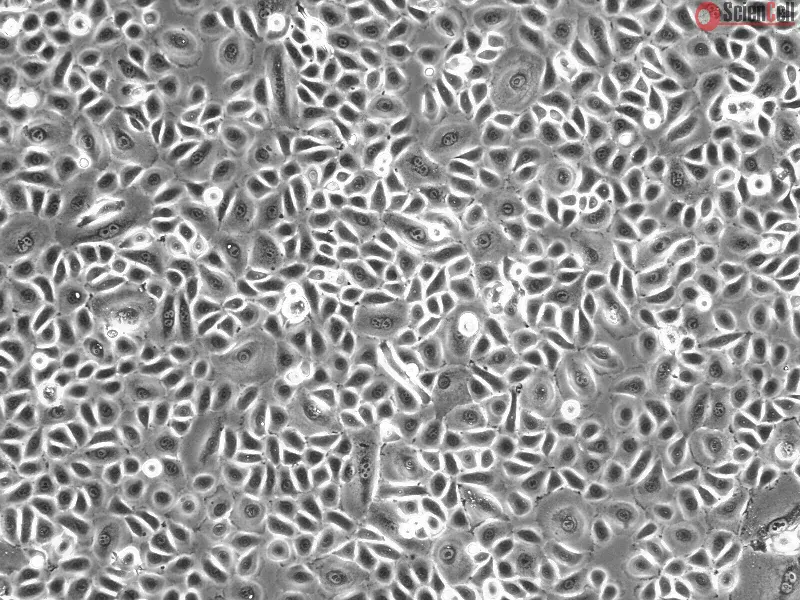
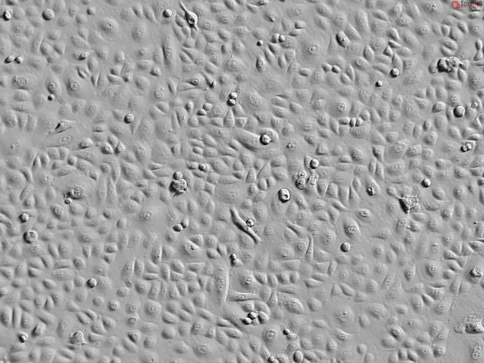
Cell culture models of the cornea are continually developed to replace the isolated animal cornea for transcorneal drug absorption studies. The aim of this study was to determine and compare epithelial tightness and permeability of currently available corneal cell culture models to avoid interlaboratory variability and to assess their usefulness for in-vitro permeation studies. Pure epithelial cell culture models (CEPI, SIRC and HCE-T cell lines), primary cultures of human corneal epithelium (HCEpiC) and the two commercially available models (RHC and Epiocular), as well as organotypic human cornea constructs (HCC, HCC-HCE-T), were investigated and data were compared with those obtained from the excised bovine cornea. Barrier properties were assessed by measurements of transepithelial electrical resistance (TEER) and permeability of three passively absorbed substances (mannitol, testosterone and timolol maleate) with different physico-chemical properties. TEER experiments revealed weak barrier functions for all of the investigated epithelial models (Less
Purpose: The aim of this study was to determine the anti-inflammatory effects of besifloxacin, a novel fluoroquinolone under clinical evaluation for the treatment of opht... More
Purpose: The aim of this study was to determine the anti-inflammatory effects of besifloxacin, a novel fluoroquinolone under clinical evaluation for the treatment of ophthalmic infections, in human corneal epithelial cells (HCEpiC). Methods: Cytokine expression in primary HCEpiC was stimulated by interleukin-1beta (IL-1beta), and Luminex technology was used to determine the effect of besifloxacin on IL-1beta-induced cytokine release. Effect of besifloxacin on nuclear factor kappa B (NFkappaB), and mitogen-activated protein kinase (MAPK) was assessed by measuring inhibitory kappa B protein (IkappaB) degradation, NFkappaB nuclear translocation, and MAPK phosphorylation by Western blotting. Moxifloxacin, a marketed fluoroquinolone, was used as the control. Anti-inflammatory efficacy of besifloxacin was also evaluated in rabbits infected with methicillin-resistant Staphylococcus aureus (MRSA). Results: Stimulation of HCEpiC with IL-1beta increased release of 12 of the 29 cytokines measured. Besifloxacin significantly inhibited IL-1beta-induced cytokine release in a dose-dependent manner, with a comparable (IL-8) or better (G-CSF, GM-CSF, IL-6, MCP-1, MIP-1beta, TGF-alpha, and TNF-alpha) efficacy compared to moxifloxacin. A significant inhibitory effect of besifloxacin was observed at 1 or 10 microg/ml. Besifloxacin inhibited IkappaB degradation, NFkappaB nuclear translocation, and activation of p38 and JNK MAPKs. Based on improvement of clinical score, besifloxacin showed statistically significant anti-inflammatory effect compared to saline treatment. Conclusions: Besifloxacin acts as an anti-inflammatory agent in corneal epithelial cells in vitro, by inhibiting the NFkappaB and MAPK pathways. Besifloxacin also exhibits anti-inflammatory efficacy in vivo. The anti-inflammatory attribute may enhance its efficacy in the treatment of ocular infections with an inflammatory component and warrants further investigation. Less
The proinflammatory cytokine interleukin-20 (IL-20) may exert the majority of its activity in the skin. We examined the effect of various treatments including several for... More
The proinflammatory cytokine interleukin-20 (IL-20) may exert the majority of its activity in the skin. We examined the effect of various treatments including several forms of phototherapy on IL-20 expression using cultured normal human epithelial keratinocytes (NHEK). Broadband UVB light, recombinant (r) IL-1 and rIL-8 increased, while hydrocortisone reduced, NHEK supernatant IL-20 levels. Elevation of NHEK IL-20 mRNA and maximal supernatant IL-20 levels occurred with a UVB light dose (40 mJ cm(-2)) that reduced cell viability by approximately 50%. While this UVB light dose also elevated supernatant IL-1 alpha and IL-8 levels, antibody neutralization studies indicated that neither of these cytokines was directly responsible for this increase in IL-20 expression. However, the elevation in IL-20 levels was fully inhibited by the p38 mitogen-activated protein kinase (MAPK) inhibitor SB-203580, suggesting involvement of this stress signaling pathway in this UVB light response. Photodynamic therapy (PDT) with the photosensitizer lemuteporfin, UVA light, cisplatin, lipopolysaccharide (LPS), tumor necrosis factor-alpha (TNF-alpha) or recombinant interferon-gamma (rIFN-gamma) either had little effect or decreased NHEK supernatant IL-20 levels. Reduced IL-20 levels paralleled the cytotoxic actions of PDT, UVA light or cisplatin and the antiproliferative effect of rIFN-gamma. Neither rIL-20 supplementation nor anti-IL-20 antibody treatments affected cell viability indicating that soluble IL-20 did not affect the short-term survival of UVB light-irradiated NHEK. Stimulation of IL-20 expression in keratinocytes by UVB light suggests that this cytokine might participate in skin responses to this ever-present environmental factor and potentially has a role in UV light-associated dermatoses. Less


,-1-mg-ml--2.jpg)