The inflammatory process seen in inflammatory bowel disease (IBD) is due to excess production of pro-inflammatory cytokines interleukin-1 (IL-1), IL-6, tumor necrosis fac... More
The inflammatory process seen in inflammatory bowel disease (IBD) is due to excess production of pro-inflammatory cytokines interleukin-1 (IL-1), IL-6, tumor necrosis factor-α (TNF-α), interferons (IFNs), macrophage migration inhibitory factor (MIF), HMGB1 (high mobility group B1) and possibly, a reduction in anti-inflammatory cytokines IL-10, IL-4, and transforming growth factor-β (TGF-β). These pro-inflammatory molecules lead to increased production of reactive oxygen species (ROS) including nitric oxide resulting in target tissue damage. I propose that inadequate production of inflammation resolving molecules lipoxins, resolvins, protectins, maresins and nitrolipids that suppress inflammation, ROS production, enhance wound healing and have cytoprotective properties results in inappropriate inflammation, delay in healing/repair process and so target tissue/organ damage continues in IBD. Hence, suggested therapeutic approach could include administration of stable synthetic analogues of lipoxins, resolvins, protectins, maresins and nitrolipids. This implies that measuring urine, stool and plasma levels of lipoxins, resolvins, protectins, maresins and nitrolipids may be used to detect the onset, progression and response to treatment of IBD. Less
The study of host-microbiota interactions in humans is largely limited to identifying associations between microbial communities and host phenotypes. While these studies ... More
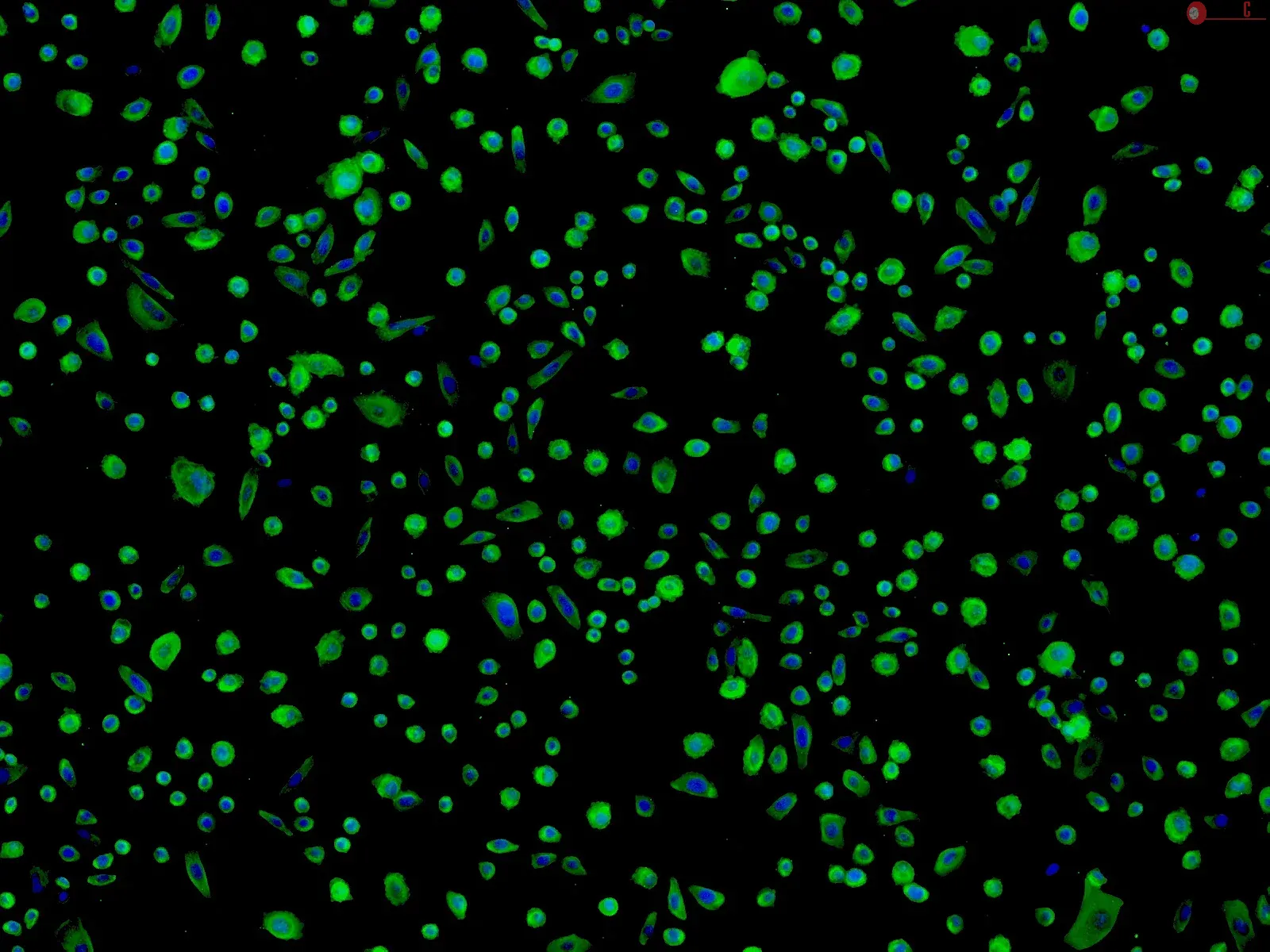
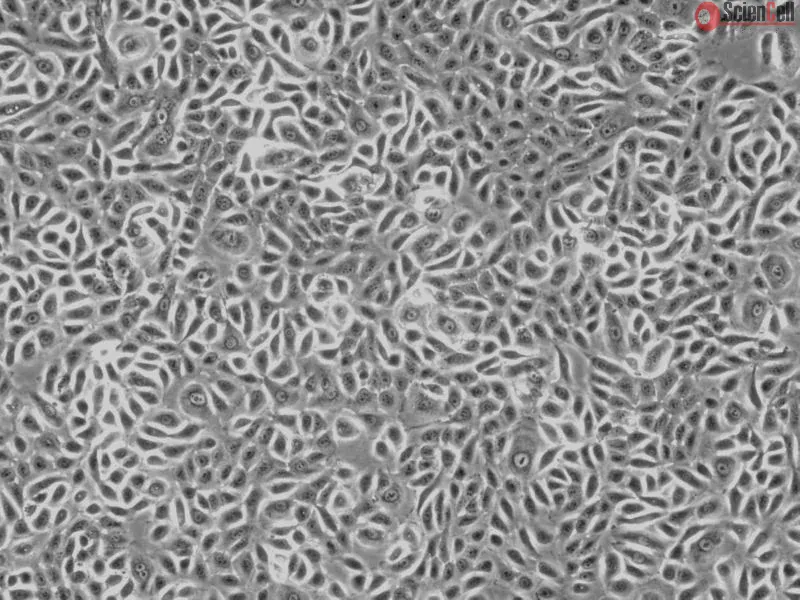
The study of host-microbiota interactions in humans is largely limited to identifying associations between microbial communities and host phenotypes. While these studies have generated important insights on the links between the microbiota and human disease, the assessment of cause-and-effect relationships has been challenging. Although this relationship can be studied in germfree mice, this system is costly, and it is difficult to accurately account for the effects of host genotypic variation and environmental effects seen in humans. Here, we have developed a novel approach to directly investigate the transcriptional changes induced by live microbial communities on human colonic epithelial cells and how these changes are modulated by host genotype. This method is easily scalable to large numbers of host genetic backgrounds and diverse microbiota and can be utilized to elucidate the mechanisms of host-microbiota interactions. Future extensions may also include colonic organoid cultures. Less
Autophagy, a type II programmed cell death, is essential for cell survival under stress, e.g. lung injury, and bone marrow-derived mesenchymal stem cells (BM-MSCs) have g... More
Autophagy, a type II programmed cell death, is essential for cell survival under stress, e.g. lung injury, and bone marrow-derived mesenchymal stem cells (BM-MSCs) have great potential for cell therapy. However, the mechanisms underlying the BM-MSC activation of autophagy to provide a therapeutic effect in ischaemia/reperfusion-induced lung injury (IRI) remain unclear. Thus, we investigate the activation of autophagy in IRI following transplantation with BM-MSCs. Seventy mice were pre-treated with BM-MSCs before they underwent lung IRI surgery in vivo. Human pulmonary micro-vascular endothelial cells (HPMVECs) were pre-conditioned with BM-MSCs by oxygen-glucose deprivation/reoxygenation (OGD) in vitro. Expression markers for autophagy and the phosphoinositide 3-kinase/protein kinase B (PI3K/Akt) signalling pathway were analysed. In IRI-treated mice, administration of BM-MSCs significantly attenuated lung injury and inflammation, and increased the level of autophagy. In OGD-treated HPMVECs, co-culture with BM-MSCs attenuated endothelial permeability by decreasing the level of cell death and enhanced autophagic activation. Moreover, administration of BM-MSCs decreased the level of PI3K class I and p-Akt while the expression of PI3K class III was increased. Finally, BM-MSCs-induced autophagic activity was prevented using the inhibitor LY294002. Administration of BM-MSCs attenuated lung injury by improving the autophagy level via the PI3K/Akt signalling pathway. These findings provide further understanding of the mechanisms related to BM-MSCs and will help to develop new cell-based therapeutic strategies in lung injury. Less
AIM: To investigate the effect of metformin on silibinin-induced apoptosis in human colorectal cancer (COLO 205) cells. METHODS: MTT assays were performed to quantify cel... More
AIM: To investigate the effect of metformin on silibinin-induced apoptosis in human colorectal cancer (COLO 205) cells. METHODS: MTT assays were performed to quantify cell viability. Western blot assays were applied to identify the expression of signaling proteins. RESULTS: The combined treatment of COLO 205 cells with metformin and silibinin decreased cell survival at a dose insufficient to influence the non-malignant cells [Human colonic epithelial cells (HCoEpiC)]. Silibinin and metformin increased phosphatase and tensin homolog and 5’-adenosine monophosphate-activated protein kinase expression in COLO 205 cells and inhibited the phosphorylation of mammol/Lalian target of rapamycin. This combined treatment resulted in an increase in the expression of activated caspase 3 and apoptosis inducing factor, indicating apoptosis. CONCLUSION: The combined treatment of human colorectal cancer cells with silibinin and metformin may induce apoptosis at a dose that does not affect HCoEpiC. This finding reveals a potential therapeutic strategy for the treatment of colorectal cancer. Keywords: Silibinin, Metformin, COLO 205, Mammol/Lalian target of rapamycin, Apoptosis, Colorectal cancer Less
Oxaliplatin is an important drug in the chemotherapy of colorectal carcinoma, but its toxicity, especially dose-related neurosensory toxicity, is not well tolerated. In t... More
Oxaliplatin is an important drug in the chemotherapy of colorectal carcinoma, but its toxicity, especially dose-related neurosensory toxicity, is not well tolerated. In this study, we investigated whether honokiol could augment the anti-tumor effect of oxaliplatin in colon cancer HT-29 cells in vitro and whether honokiol could be used with oxaliplatin to decrease oxaliplatin dose. We used the normal colon cells, human colonic epithelial cells (HCoEpiCs) as control cells. Cell proliferation, apoptosis, prostaglandin E2 (PGE2) and vascular endothelial growth factor (VEGF) levels were also investigated. Expression levels of cyclo-oxygenase 2 (COX-2), VEGF, AKT/p-AKT, extracellular signal-related kinase (ERK)1/2/p-ERK1/2, nuclear factor kappa B (NF-κB) P65/p-P65, and caspase-3 were measured. Honokiol or oxaliplatin suppressed the proliferation of HT-29 cells in a concentration-dependent manner, but only high concentrations of honokiol would suppress the proliferation of HCoEpiCs. HT-29 cells were more sensitive to oxaliplatin treatment in the presence of honokiol. Oxaliplatin combined with honokiol improved the apoptosis rate of HT-29 cell and reduced PGE2 and VEGF secretion levels. Expression levels of COX-2 and VEGF protein and phosphorylation of AKT, ERK1/2, and NF-κB P65 were also inhibited. Caspase-3 levels were upregulated after honokiol treatment. Therefore, honokiol can be used in combination with oxaliplatin in the chemotherapy of colon cancer. This combination allows a reduction in oxaliplatin dose, and thereby reduces its adverse effects. It may also enhance the chemotherapeutic effect of oxaliplatin for this disease. Less
In colon cancer, a highly aggressive disease, progression through the malignant sequence is accompanied by increasingly numerous chromosomal rearrangements. To colonize t... More
In colon cancer, a highly aggressive disease, progression through the malignant sequence is accompanied by increasingly numerous chromosomal rearrangements. To colonize target organs, invasive cells cross several tissues of various elastic moduli. Whether soft tissue increases malignancy or in contrast limits invasive colon cell spreading remains an open question. Using polyelectrolyte multilayer films mimicking microenvironments of various elastic moduli, we revealed that human SW480 colon cancer cells displayed increasing frequency in chromosomal segregation abnormalities when cultured on substrates with decreasing stiffness. Our results show that, although decreasing stiffness correlates with increased cell lethality, a significant proportion of SW480 cancer cells did escape from the very soft substrates, even when bearing abnormal chromosome segregation, achieve mitosis and undergo a new cycle of replication in contrast to human colonic HCoEpiC cells which died on soft substrates. This observation opens the possibility that the ability of cancer cells to overcome defects in chromosome segregation on very soft substrates could contribute to increasing chromosomal rearrangements and tumor cell aggressiveness. Less


,-1-mg-ml--2.jpg)