The choroid plexus is located in the ventricles of the brain where cerebrospinal fluid (CSF) is constantly produced. It consists of a network of capillaries enclosed by a single layer of epithelial cells that together form the blood-CSF barrier. Choroid plexus epithelial cells (CPEpiC) play a central role in restricting the passage of molecules and ions between the brain and CSF by the junctional complexes between neighboring cells. They also have a special cellular structure, and channel and transporter protein expression well-adapted for CSF secretion. A previous study further demonstrated that CPEpiC respond to ischemic and traumatic insults in the central nervous system by synthesizing and secreting various growth factors and peptides with trophic benefits that work through autocrine and paracrine mechanisms to facilitate recovery. CPEpiC cultures serve as an excellent model to study the exchange processes across blood-CSF barrier, CSF homeostasis,
and more.
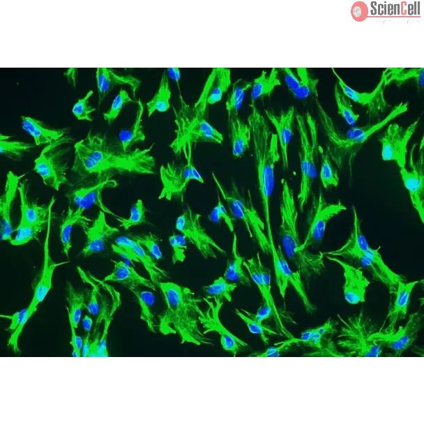
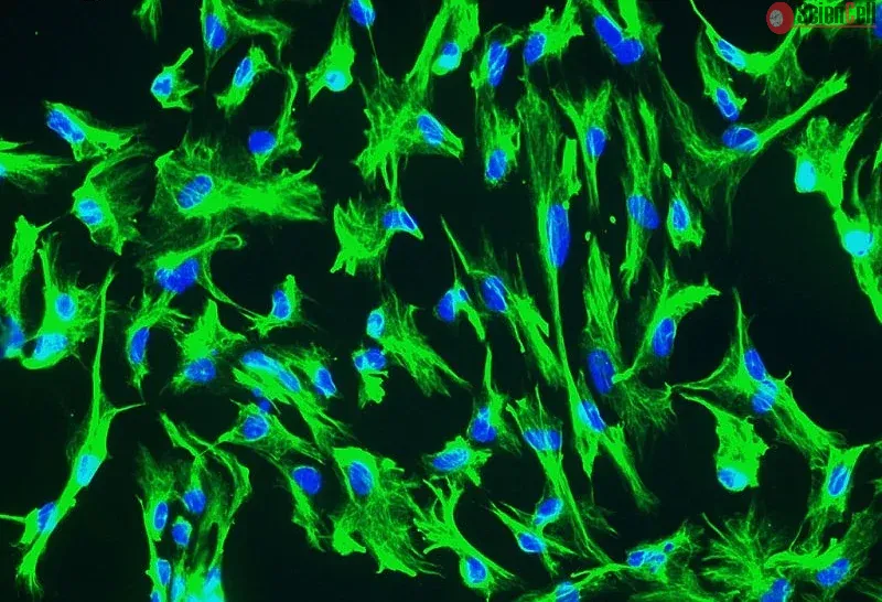
HCPEpiC from ScienCell Research Laboratories are isolated from human choroid plexus. HCPEpiC are cryopreserved after purification and delivered frozen. Each vial contains >5 x 10⁵ cells in 1 ml volume. HCPEpiC are characterized by immunofluorescence with antibodies specific to cytokeratin-18 and/or cytokeratin-19. HCPEpiC are negative for HIV-1, HBV, HCV, mycoplasma, bacteria, yeast, and fungi. HCPEpiC are guaranteed to further expand for 5 population doublings under the conditions provided by ScienCell Research Laboratories.
Recommended Medium
It is recommended to use Epithelial Cell Medium (EpiCM, Cat. #4101) for culturing HCPEpiC in vitro.
Background: The content and composition of cerebrospinal fluid (CSF) is determined in large part by the choroid plexus (CP) and specifically, a specialized epithelial cel... More
Background: The content and composition of cerebrospinal fluid (CSF) is determined in large part by the choroid plexus (CP) and specifically, a specialized epithelial cell (CPe) layer that responds to, synthesizes, and transports peptide hormones into and out of CSF. Together with ventricular ependymal cells, these CPe relay homeostatic signals throughout the central nervous system (CNS) and regulate CSF hydrodynamics. One new candidate signal is augurin, a newly recognized 14 kDa protein that is encoded by esophageal cancer related gene-4 (Ecrg4), a putative tumor suppressor gene whose presence and function in normal tissues remains unexplored and enigmatic. The aim of this study was to explore whether Ecrg4 and its product augurin, can be implicated in CNS development and the response to CNS injury. Methods: Ecrg4 gene expression in CNS and peripheral tissues was studied by in situ hybridization and quantitative RT-PCR. Augurin, the protein encoded by Ecrg4, was detected by immunoblotting, immunohistochemistry and ELISA. The biological consequence of augurin over-expression was studied in a cortical stab model of rat CNS injury by intra-cerebro-ventricular injection of an adenovirus vector containing the Ecrg4 cDNA. The biological consequences of reduced augurin expression were evaluated by characterizing the CNS phenotype caused by Ecrg4 gene knockdown in developing zebrafish embryos. Results: Gene expression and immunohistochemical analyses revealed that, the CP is a major source of Ecrg4 in the CNS and that Ecrg4 mRNA is predominantly localized to choroid plexus epithelial (CPe), ventricular and central canal cells of the spinal cord. After a stab injury into the brain however, both augurin staining and Ecrg4 gene expression decreased precipitously. If the loss of augurin was circumvented by over-expressing Ecrg4 in vivo, BrdU incorporation by cells in the subependymal zone decreased. Inversely, gene knockdown of Ecrg4 in developing zebrafish embryos caused increased proliferation of GFAP-positive cells and induced a dose-dependent hydrocephalus-like phenotype that could be rescued by co-injection of antisense morpholinos with Ecrg4 mRNA. Conclusion: An unusually elevated expression of the Ecrg4 gene in the CP implies that its product, augurin, plays a role in CP-CSF-CNS function. The results are all consistent with a model whereby an injury-induced decrease in augurin dysinhibits target cells at the ependymal-subependymal interface. We speculate that the ability of CP and ependymal epithelium to alter the progenitor cell response to CNS injury may be mediated, in part by Ecrg4. If so, the canonic control of its promoter by DNA methylation may implicate epigenetic mechanisms in neuroprogenitor fate and function in the CNS. Less
Approximately 500 million people worldwide are chronically infected with the hepatitis B virus (HBV) or hepatitis C virus (HCV), and are therefore at an increased risk fo... More
Approximately 500 million people worldwide are chronically infected with the hepatitis B virus (HBV) or hepatitis C virus (HCV), and are therefore at an increased risk for developing fatal liver diseases such as cirrhosis and hepatocellular carcinoma. The intracellular antiviral responses induced by interferon (IFN)-α/-β and/or IFN-γ play critical roles in the pathogenesis of HBV and HCV infection, and the function of IFN-λ in the host immune response to these viruses is beginning to be revealed. A better understanding of how IFN-λ influences HBV or HCV persistence is not only important for understanding the mechanisms of chronic virus infection, but also may lead to new approaches for improved antiviral therapies. Less
ScienCell Research Laboratories (SRL) takes pride in being a resource for researchers all over the world. The publications listed here are not meant as an endorsement or confirmation of the reliability of the products.

,-1-mg-ml--2.jpg)