Human cardiac fibroblasts (HCFs) have various voltage-dependent K(+) channels (VDKCs) that can induce apoptosis. Hydrogen peroxide (H2O2) modulates VDKCs and induces oxid... More
Human cardiac fibroblasts (HCFs) have various voltage-dependent K(+) channels (VDKCs) that can induce apoptosis. Hydrogen peroxide (H2O2) modulates VDKCs and induces oxidative stress, which is the main contributor to cardiac injury and cardiac remodeling. We investigated whether H2O2 could modulate VDKCs in HCFs and induce cell injury through this process. In whole-cell mode patch-clamp recordings, application of H2O2 stimulated Ca(2+)-activated K(+) (KCa) currents but not delayed rectifier K(+) or transient outward K(+) currents, all of which are VDKCs. H2O2-stimulated KCa currents were blocked by iberiotoxin (IbTX, a large conductance KCa blocker). The H2O2-stimulating effect on large-conductance KCa (BKCa) currents was also blocked by KT5823 (a protein kinase G inhibitor) and 1 H-[1, 2, 4] oxadiazolo-[4, 3-a] quinoxalin-1-one (ODQ, a soluble guanylate cyclase inhibitor). In addition, 8-bromo-cyclic guanosine 3', 5'-monophosphate (8-Br-cGMP) stimulated BKCa currents. In contrast, KT5720 and H-89 (protein kinase A inhibitors) did not block the H2O2-stimulating effect on BKCa currents. Using RT-PCR and western blot analysis, three subtypes of KCa channels were detected in HCFs: BKCa channels, small-conductance KCa (SKCa) channels, and intermediate-conductance KCa (IKCa) channels. In the annexin V/propidium iodide assay, apoptotic changes in HCFs increased in response to H2O2, but IbTX decreased H2O2-induced apoptosis. These data suggest that among the VDKCs of HCFs, H2O2 only enhances BKCa currents through the protein kinase G pathway but not the protein kinase A pathway, and is involved in cell injury through BKCa channels. Keywords: Ca2+-activated K+ channels; Human cardiac fi broblasts; Hydrogen peroxide; K+ currents; Protein kinase. Less
Autophagy, a type II programmed cell death, is essential for cell survival under stress, e.g. lung injury, and bone marrow-derived mesenchymal stem cells (BM-MSCs) have g... More
Autophagy, a type II programmed cell death, is essential for cell survival under stress, e.g. lung injury, and bone marrow-derived mesenchymal stem cells (BM-MSCs) have great potential for cell therapy. However, the mechanisms underlying the BM-MSC activation of autophagy to provide a therapeutic effect in ischaemia/reperfusion-induced lung injury (IRI) remain unclear. Thus, we investigate the activation of autophagy in IRI following transplantation with BM-MSCs. Seventy mice were pre-treated with BM-MSCs before they underwent lung IRI surgery in vivo. Human pulmonary micro-vascular endothelial cells (HPMVECs) were pre-conditioned with BM-MSCs by oxygen-glucose deprivation/reoxygenation (OGD) in vitro. Expression markers for autophagy and the phosphoinositide 3-kinase/protein kinase B (PI3K/Akt) signalling pathway were analysed. In IRI-treated mice, administration of BM-MSCs significantly attenuated lung injury and inflammation, and increased the level of autophagy. In OGD-treated HPMVECs, co-culture with BM-MSCs attenuated endothelial permeability by decreasing the level of cell death and enhanced autophagic activation. Moreover, administration of BM-MSCs decreased the level of PI3K class I and p-Akt while the expression of PI3K class III was increased. Finally, BM-MSCs-induced autophagic activity was prevented using the inhibitor LY294002. Administration of BM-MSCs attenuated lung injury by improving the autophagy level via the PI3K/Akt signalling pathway. These findings provide further understanding of the mechanisms related to BM-MSCs and will help to develop new cell-based therapeutic strategies in lung injury. Less
Aims Epidemiological and interventional studies have suggested a protective role for vitamin D in cardiovascular disease, and basic research has implicated vitamin D as a... More
Aims Epidemiological and interventional studies have suggested a protective role for vitamin D in cardiovascular disease, and basic research has implicated vitamin D as a potential inhibitor of fibrosis in a number of organ systems; yet little is known regarding direct effects of vitamin D on human cardiac cells. Given the critical role of fibrotic responses in end stage cardiac disease, we examined the effect of active vitamin D treatment on fibrotic responses in primary human adult ventricular cardiac fibroblasts (HCF-av), and investigated the relationship between circulating vitamin D (25(OH)D3) and cardiac fibrosis in human myocardial samples. Methods and Results Interstitial cardiac fibrosis in end stage HF was evaluated by image analysis of picrosirius red stained myocardial sections. Serum 25(OH)D3 levels were assayed using mass spectrometry. Commercially available HCF-av were treated with transforming growth factor (TGF)β1 to induce activation, in the presence or absence of active vitamin D (1,25(OH)2D3). Functional responses of fibroblasts were analyzed by in vitro collagen gel contraction assay. 1,25(OH)2D3 treatment significantly inhibited TGFβ1-mediated cell contraction, and confocal imaging demonstrated reduced stress fiber formation in the presence of 1,25(OH)2D3. Treatment with 1,25(OH)2D3 reduced alpha-smooth muscle actin expression to control levels and inhibited SMAD2 phosphorylation. Conclusions Our results demonstrate that active vitamin D can prevent TGFβ1-mediated biochemical and functional pro-fibrotic changes in human primary cardiac fibroblasts. An inverse relationship between vitamin D status and cardiac fibrosis in end stage heart failure was observed. Collectively, our data support an inhibitory role for vitamin D in cardiac fibrosis. Less
Heart failure (HF) prevention strategies require biomarkers that identify disease manifestation. Increases in B-type natriuretic peptide (BNP) correlate with increased ri... More
Heart failure (HF) prevention strategies require biomarkers that identify disease manifestation. Increases in B-type natriuretic peptide (BNP) correlate with increased risk of cardiovascular events and HF development. We hypothesize that coronary sinus serum from a high BNP hypertensive population reflects an active pathological process and can be used for biomarker exploration. Our aim was to discover differentially expressed disease-associated proteins that identify patients with ventricular dysfunction and HF. Coronary sinus serum from 11 asymptomatic, hypertensive patients underwent quantitative differential protein expression analysis by 2-dimensional difference gel electrophoresis. Proteins were identified using mass spectrometry and then studied by enzyme-linked immunosorbent assay in sera from 40 asymptomatic, hypertensive patients and 105 patients across the spectrum of ventricular dysfunction (32 asymptomatic left ventricular diastolic dysfunction, 26 diastolic HF, and 47 systolic HF patients). Leucine-rich α2-glycoprotein (LRG) was consistently overexpressed in high BNP serum. LRG levels correlate significantly with BNP in hypertensive, asymptomatic left ventricular diastolic dysfunction, diastolic HF, and systolic HF patient groups (P≤0.05). LRG levels were able to identify HF independent of BNP. LRG correlates with coronary sinus serum levels of tumor necrosis factor-α (P=0.009) and interleukin-6 (P=0.021). LRG is expressed in myocardial tissue and correlates with transforming growth factor-βR1 (P<0.001) and α-smooth muscle actin (P=0.025) expression. LRG was identified as a serum biomarker that accurately identifies patients with HF. Multivariable modeling confirmed that LRG is a stronger identifier of HF than BNP and this is independent of age, sex, creatinine, ischemia, β-blocker therapy, and BNP. Less
Ca(2+) signaling pathways are well studied in cardiac myocytes, but not in cardiac fibroblasts. The aim of the present study is to characterize Ca(2+) signaling pathways ... More
Ca(2+) signaling pathways are well studied in cardiac myocytes, but not in cardiac fibroblasts. The aim of the present study is to characterize Ca(2+) signaling pathways in cultured human cardiac fibroblasts using confocal scanning microscope and RT-PCR techniques. It was found that spontaneous intracellular Ca(2+) (Ca(i) (2+)) oscillations were present in about 29% of human cardiac fibroblasts, and the number of cells with Ca(i) (2+) oscillations was increased to 57.3% by application of 3% fetal bovine serum. Ca(i) (2+) oscillations were dependent on Ca(2+) entry. Ca(i) (2+) oscillations were abolished by the store-operated Ca(2+) (SOC) entry channel blocker La(3+), the phospholipase C inhibitor U-73122, and the inositol trisphosphate receptors (IP3Rs) inhibitor 2-aminoethoxydiphenyl borate, but not by ryanodine. The IP3R agonist thimerosal enhanced Ca(i) (2+) oscillations. Inhibition of plasma membrane Ca(2+) pump (PMCA) and Na(+)-Ca(2+) exchanger (NCX) also suppressed Ca(i) (2+) oscillations. In addition, the frequency of Ca(i) (2+) oscillations was reduced by nifedipine, and increased by Bay K8644 in cells with spontaneous Ca(2+) oscillations. RT-PCR revealed that mRNAs for IP3R1-3, SERCA1-3, Ca(V)1.2, NCX3, PMCA1,3,4, TRPC1,3,4,6, STIM1, and Orai1-3, were readily detectable, but not RyRs. Our results demonstrate for the first time that spontaneous Ca(i) (2+) oscillations are present in cultured human cardiac fibroblasts and are regulated by multiple Ca(2+) pathways, which are not identical to those of the well-studied contractile cardiomyocytes. This study provides a base for future investigations into how Ca(2+) signals regulate biological activity in human cardiac fibroblasts and cardiac remodeling under pathological conditions. Less
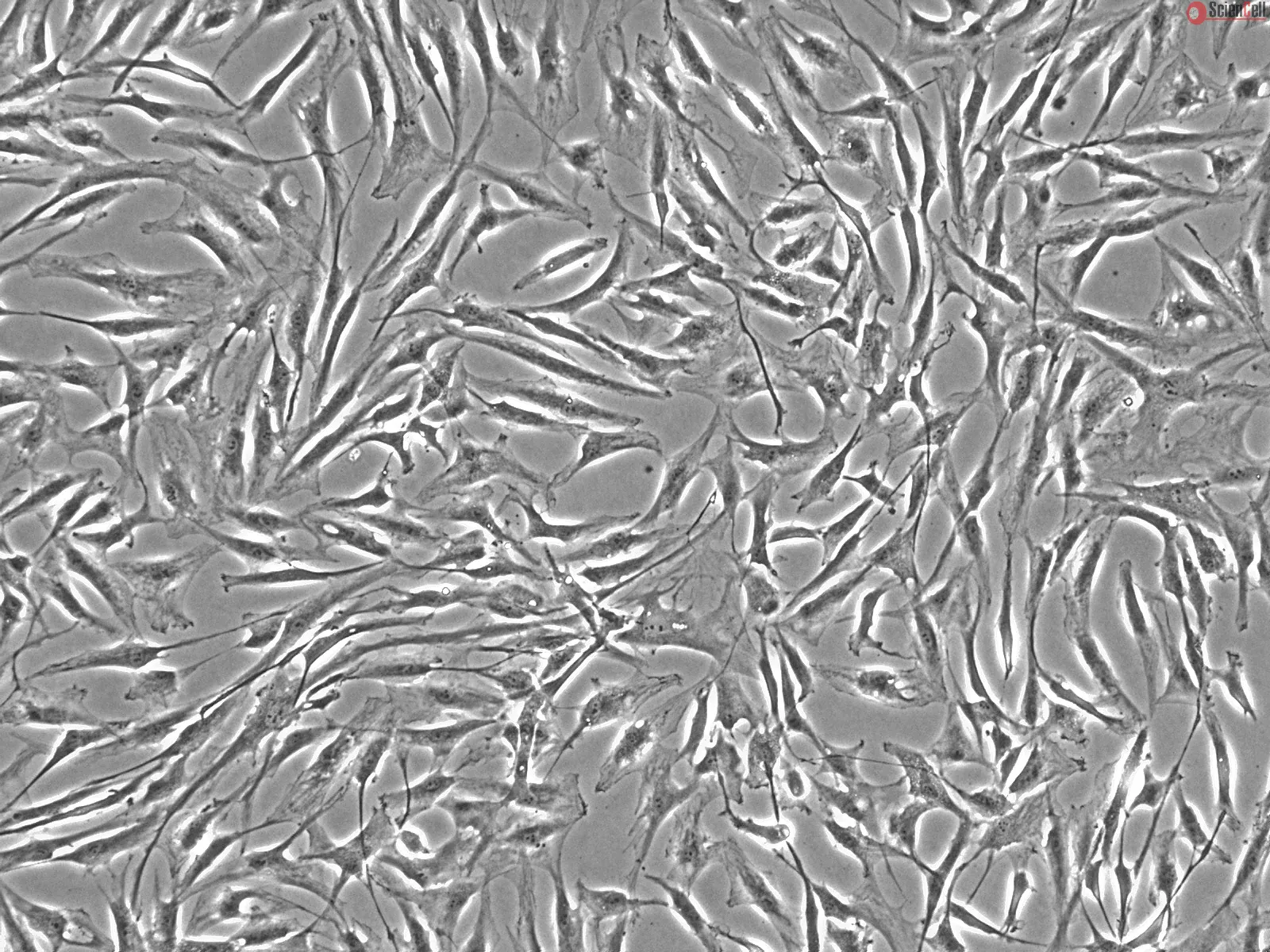
Background: Although fibroblast-to-myocyte electrical coupling is experimentally suggested, electrophysiology of cardiac fibroblasts is not as well established as contrac... More
Background: Although fibroblast-to-myocyte electrical coupling is experimentally suggested, electrophysiology of cardiac fibroblasts is not as well established as contractile cardiac myocytes. The present study was therefore designed to characterize ion channels in cultured human cardiac fibroblasts. Less
Aldosterone is known to regulate electrolyte homeostasis, but it may also contribute to other processes, including the maladaptive remodeling of postinfarct hearts. Becau... More
Aldosterone is known to regulate electrolyte homeostasis, but it may also contribute to other processes, including the maladaptive remodeling of postinfarct hearts. Because aldosterone has been implicated in the stimulation of collagen production in the heart, we investigated whether it would also affect elastin deposition in cultures of human cardiac fibroblasts. We first demonstrated that treatment with 1 to 50 nmol/L aldosterone leads to a significant increase in collagen type I mRNA levels and in subsequent collagen fiber deposition. Pretreatment of cells with the mineralocorticoid receptor antagonist spironolactone, but not with the glucocorticoid receptor antagonist RU 486, inhibited collagen synthesis in aldosterone-treated cultures. Most importantly, we demonstrated that aldosterone also increases elastin mRNA levels, tropoelastin synthesis, and elastic fiber deposition in a dose-dependent manner. Strikingly, neither spironolactone nor RU 486 eliminated aldosterone-induced increases in elastin production. We further discovered that the proelastogenic effect of aldosterone involves a rapid increase in tyrosine phosphorylation of the insulin-like growth factor-I receptor and that the insulin-like growth factor-I receptor kinase inhibitor AG1024 or an anti-insulin-like growth factor-I receptor-neutralizing antibody inhibits both insulin-like growth factor-I and aldosterone-induced elastogenesis. Thus, we have demonstrated for the first time that aldosterone, which stimulates collagen production through the mineralocorticoid receptor-dependent pathway, also increases elastogenesis via a parallel mineralocorticoid receptor-independent pathway involving I insulin-like growth factor-I receptor signaling. Less

,-1-mg-ml--2.jpg)