A key aim of therapy for multiple sclerosis (MS) is to promote the regeneration of oligodendrocytes and remyelination in the central nervous system (CNS). The present stu... More
A key aim of therapy for multiple sclerosis (MS) is to promote the regeneration of oligodendrocytes and remyelination in the central nervous system (CNS). The present study provides evidence that the vitamin K-dependent protein growth arrest specific 6 (Gas6) promotes such repair in in vitro cultures of mouse optic nerve and cerebellum. We first determined expression of Gas6 and TAM (Tyro3, Axl, Mer) receptors in the mouse CNS, with all three TAM receptors increasing in expression through postnatal development, reaching maximal levels in the adult. Treatment of cultured mouse optic nerves with Gas6 resulted in significant increases in oligodendrocyte numbers as well as expression of myelin basic protein (MBP). Gas6 stimulation also resulted in activation of STAT3 in optic nerves as well as downregulation of multiple genes involved in MS development, including matrix metalloproteinase-9 (MMP9), which may decrease the integrity of the blood-brain barrier and is found upregulated in MS lesions. The cytoprotective effects of Gas6 were examined in in vitro mouse cerebellar slice cultures, where lysolecithin was used to induce demyelination. Cotreatment of cerebellar slices with Gas6 significantly attenuated demyelination as determined by MBP immunostaining, and Gas6 activated Tyro3 receptor through its phosphorylation. In conclusion, these results demonstrate that Gas6/TAM signaling stimulates the generation of oligodendrocytes and increased myelin production via Tyro3 receptor in the adult CNS, including repair after demyelinating injury. Furthermore, the effects of Gas6 on STAT3 signaling and matrix MMP9 downregulation indicate potential glial cell repair and immunoregulatory roles for Gas6, indicating that Gas6-TAM signaling could be a potential therapeutic target in MS and other neuropathologies. Keywords: TAM receptors; Tyro3; multiple sclerosis; myelin; oligodendrocyte; receptor tyrosine kinase. © The Author(s) 2016. Less
Previous studies have shown that the tumor necrosis factor-related apoptosis-inducing ligand (TRAIL) has significant apoptosis-inducing activity in some glioma cell lines... More
Previous studies have shown that the tumor necrosis factor-related apoptosis-inducing ligand (TRAIL) has significant apoptosis-inducing activity in some glioma cell lines, although many lines are either moderately or completely resistant, which has limited the therapeutic applicability of this agent. Because our recent studies showed that inhibition of proteasomal function may be independently active as an apoptosis-inducing stimulus in these tumors, we investigated the sensitivity of a panel of glioma cell lines (U87, T98G, U373, A172, LN18, LN229, LNZ308, and LNZ428) to TRAIL alone and in combination with the proteasome inhibitor bortezomib. Analysis of these cell lines revealed marked differences in their sensitivity to these treatments, with two (LNZ308 and U373) of the eight cell lines revealing no significant induction of cell death in response to TRAIL alone. No correlation was found between sensitivity of cells to TRAIL and expression of TRAIL receptors DR4, DR5, and decoy receptor DcR1, caspase 8, apoptosis inhibitory proteins XIAP, survivin, Mcl-1, Bcl-2, Bcl-Xl, and cFLIP. However, TRAIL-resistant cell lines exhibited a high level of basal NF-κB activity. Bortezomib was capable of potentiating TRAIL-induced apoptosis in TRAIL-resistant cells in a caspase-dependent fashion. Bortezomib abolished p65/NF-κB DNA-binding activity, supporting the hypothesis that inhibition of the NF-κB pathway is critical for the enhancement of TRAIL sensitization in glioma cells. Moreover, knockdown of p65/NF-κB by shRNA also enhanced TRAIL-induced apoptosis, indicating that p65/NF-κB may be important in mediating TRAIL sensitivity and the effect of bortezomib in promoting TRAIL sensitization and apoptosis induction. ©2010 AACR. Less
Background: Src family kinases (SFK) collectively regulate a variety of cellular functions in many cancer types, including proliferation, invasion, motility, survival, di... More
Background: Src family kinases (SFK) collectively regulate a variety of cellular functions in many cancer types, including proliferation, invasion, motility, survival, differentiation, and angiogenesis. Although Dasatinib (BMS-354825), an ATP-competitive, small molecule tyrosine kinase inhibitor, suppresses the activity of SFKs at nanomolar concentrations, IC50 values for antiproliferative effects in glioma cell lines were well above the clinically achievable range, suggesting the need to interfere with other components of receptor-induced downstream signaling in order to achieve an optimal therapeutic effect. Materials and methods: The cytotoxic effects of combining Src and STAT3 inhibition on glioma cell lines were evaluated using assays to measure cell proliferation, apoptosis and migration. Western blotting and immunocytochemistry was used to monitor its effects on cell signaling and morphology. Results: Silencing Src and STAT3 expression each partially inhibited cell proliferation and migration. In addition, JSI-124 significantly enhanced the efficacy of dasatinib in vitro. Combination of dasatinib and JSI-124 achieved significant inhibition of migration in all cell lines, which correlated with the inhibition of Src and downstream mediators of adhesion (e.g. focal adhesion kinase). Cells exposed to dasatinib and JSI-124 exhibited morphological changes that were consistent with an upstream role for Src in regulating focal adhesion complexes. Conclusions: Targeting the Src and STAT pathways may contribute to the treatment of cancers that demonstrate increased levels of these signaling mediators, including malignant human glioma. Clinical studies in these tumor types are warranted. Keywords: Apoptosis; JSI-124; dasatinib; glioma; migration. Less
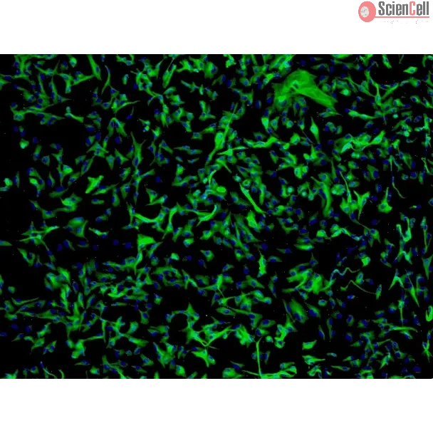
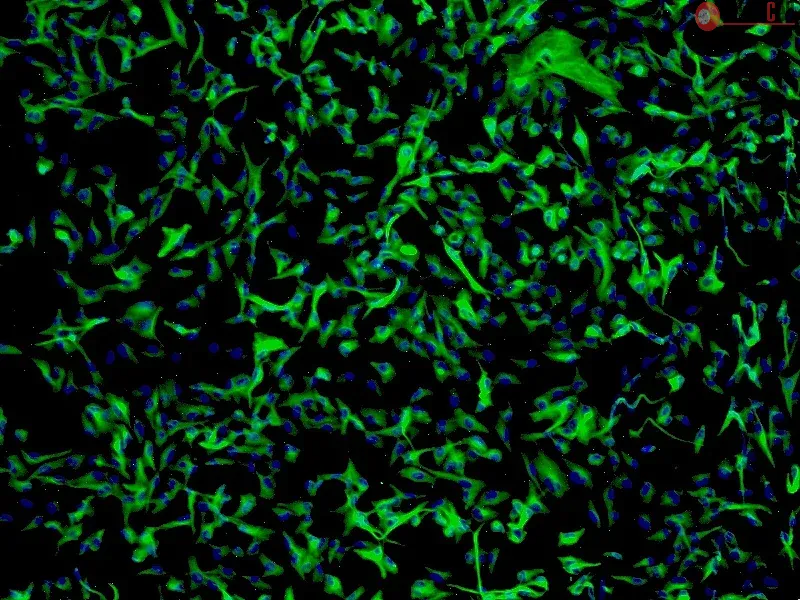
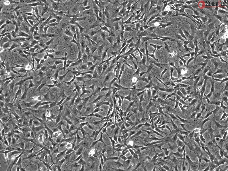
The reprogramming of human somatic cells to induced pluripotent stem (hiPS) cells enables the possibility of generating patient-specific autologous cells for regenerative... More
The reprogramming of human somatic cells to induced pluripotent stem (hiPS) cells enables the possibility of generating patient-specific autologous cells for regenerative medicine. A number of human somatic cell types have been reported to generate hiPS cells, including fibroblasts, keratinocytes and peripheral blood cells, with variable reprogramming efficiencies and kinetics. Here, we show that human astrocytes can also be reprogrammed into hiPS (ASThiPS) cells, with similar efficiencies to keratinocytes, which are currently reported to have one of the highest somatic reprogramming efficiencies. ASThiPS lines were indistinguishable from human embryonic stem (ES) cells based on the expression of pluripotent markers and the ability to differentiate into the three embryonic germ layers in vitro by embryoid body generation and in vivo by teratoma formation after injection into immunodeficient mice. Our data demonstrates that a human differentiated neural cell type can be reprogrammed to pluripotency and is consistent with the universality of the somatic reprogramming procedure. Less


,-1-mg-ml--2.jpg)
