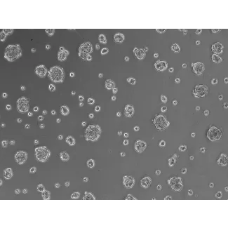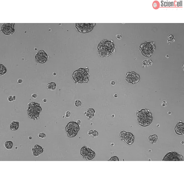
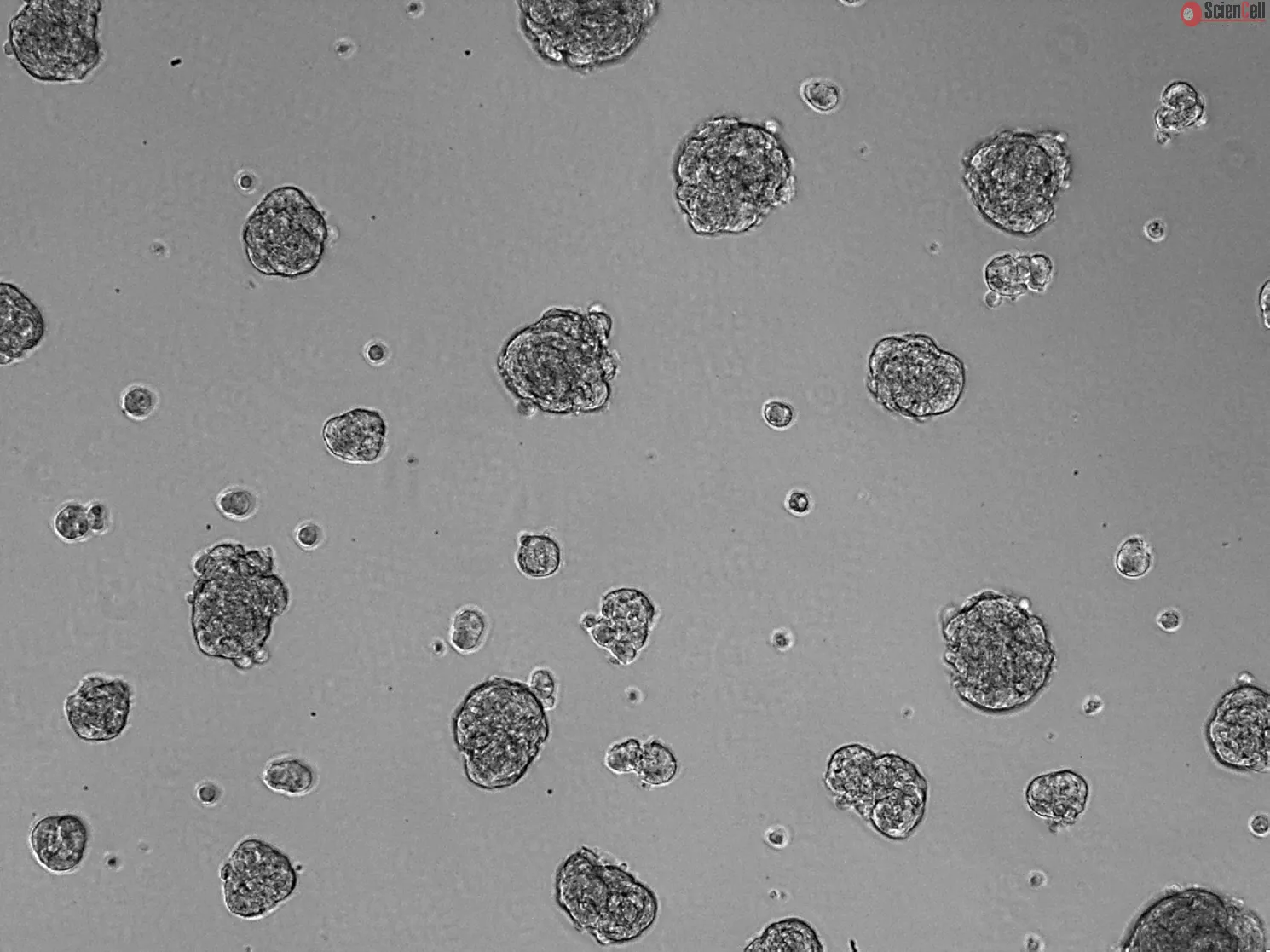
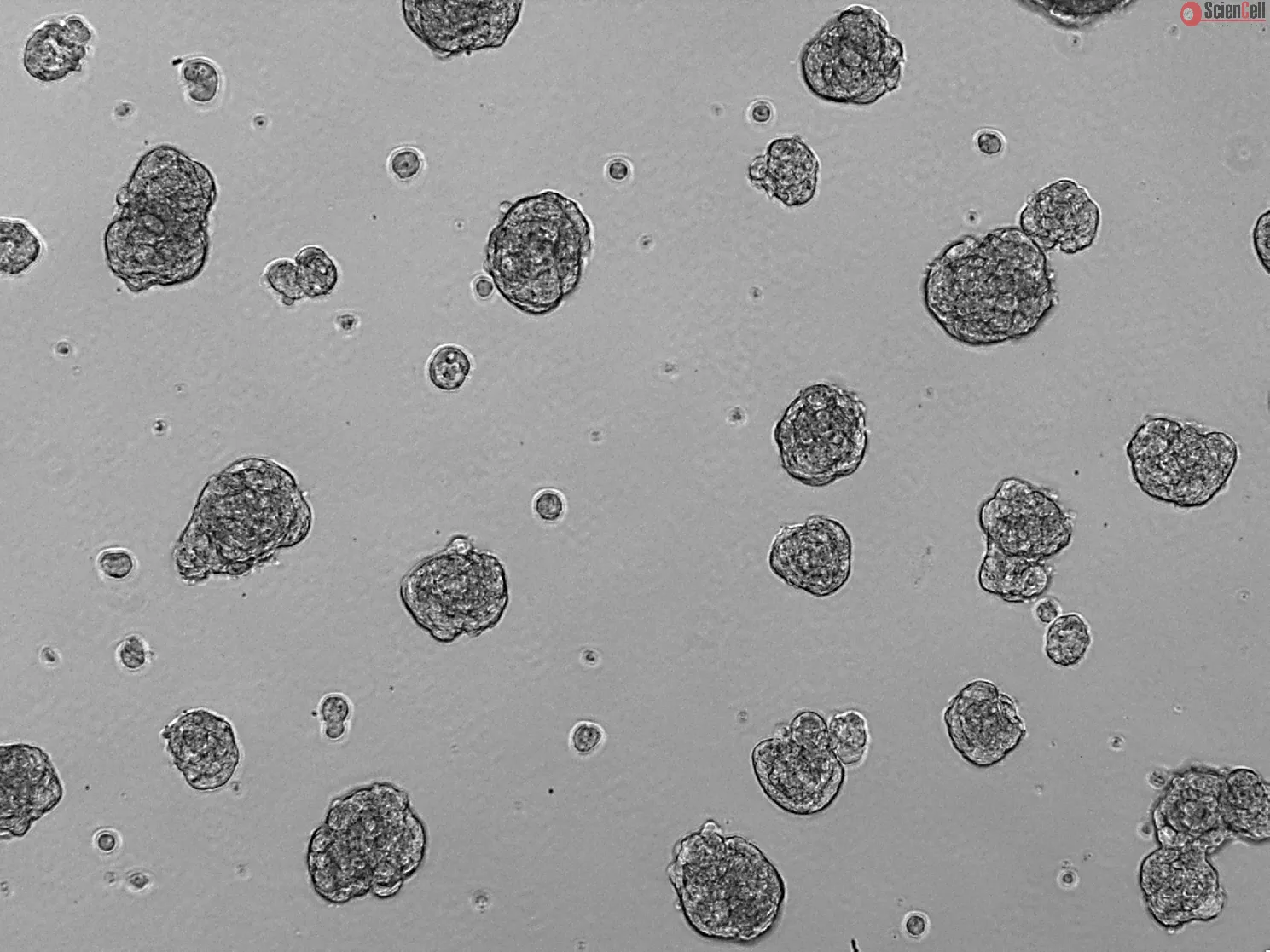
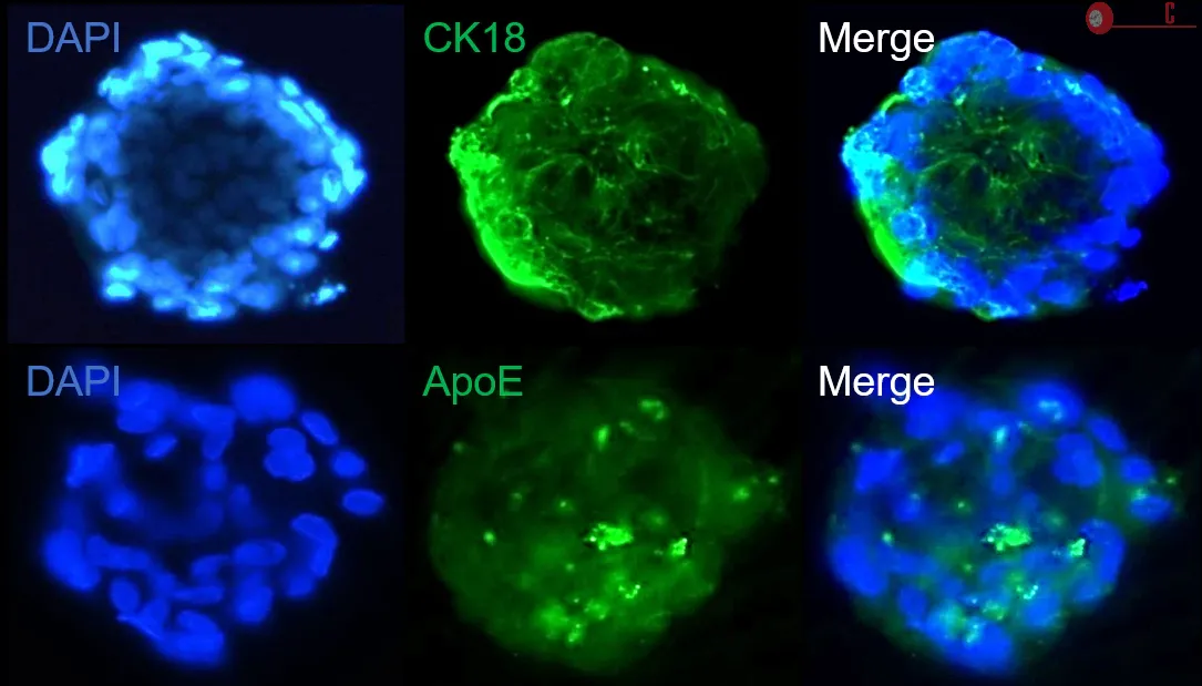
即用型3D人视网膜色素上皮细胞球状体
ScienCell的3D人视网膜色素上皮(RPE)细胞球状体表达分化上皮细胞标志物细胞角蛋白18(Cytokeratin-18),并可沉积载脂蛋白E(ApoE),后者是玻璃膜疣(drusen)的重要组成成分。该3D RPE球状体模型是体外模拟玻璃膜疣形成及研究相关疾病(如年龄相关性黄斑变性,AMD)发病机制的理想研究工具。
Age-related macular degeneration (AMD) is characterized in its early stages by the presence of extracellular deposits, known as drusen, that accumulate between the basal surface of the retinal pigmented epithelium and Bruch's membrane, an extracellular matrix complex that separates the neural retina from the capillary network in the choroid [1]. Several studies have shown that drusen contains a variety of protein and lipid components [2]. Although liver is the primary biosynthetic site for most of these molecules, retinal pigment epithelial (RPE) cells locally synthesize a number of drusen components [2]. The respective contributions of RPE-derived and plasma-derived molecules to the biogenesis of drusen, and the relevant molecular interactions leading to drusen depositions, however, have not been fully identified. One of the major limitations is that RPE cells, once isolated from the eye, tend to dedifferentiate into myofibroblasts in conventional 2D cell culture. Recent studies have shown that 3D retinal pigment epithelial cell spheroids form and maintain a well-differentiated epithelium in 3D cell culture [3]. ScienCell Research Laboratories, as a result, has developed ready-to-use 3D human retinal pigment epithelial spheroids (SP3D-HRPEpiS). 3D RPE spheroids exhibit the differentiated epithelial cell marker cytokeratin-18 and deposit apolipoprotein ApoE, a prominent drusen constituent. The 3D RPE spheroid model is an ideal way to model drusen in vitro and study the pathogenesis of related diseases, such as AMD.
Kit Components:
| 3D Cell Culture Components | ||||
|---|---|---|---|---|
| Cat # | # of vials | Product Name | Quantity | Storage |
| SP-6540 | 1 | Human Retinal Pigment Epithelial Spheroids (SP-HRPEpiS) | 1 × 10^4 spheroids | Liquid nitrogen |
| 3D-4101 | 1 | 3D-Epithelial Spheroid Medium (3D-EpiSpM) | 200 mL | 2-8 °C |
| 0004 | 1 | Fetal Bovine Serum (FBS) | 4 mL | -20 °C |
| 0583 | 1 | Penicillin/Streptomycin Solution (P/S) | 2 mL | -20 °C |
| 0343 or 0353 or 0383 | 1 | Ultra-Low Binding Culture Plates (24-, 48-, or 96- well plate) | 1 plate | RT |
年龄相关性黄斑变性(AMD)在疾病早期的典型特征是在视网膜色素上皮(RPE)基底面与Bruch膜之间形成细胞外沉积物,即玻璃膜疣(drusen)。Bruch膜是一种细胞外基质复合结构,将神经视网膜与脉络膜毛细血管网络分隔开来[1]。多项研究表明,玻璃膜疣由多种蛋白质和脂质成分组成[2]。尽管其中大多数分子主要由肝脏合成,但RPE细胞也能够在局部合成多种玻璃膜疣相关成分[2]。然而,目前RPE来源与血浆来源分子在玻璃膜疣形成中的相对贡献,以及促进玻璃膜疣沉积的分子机制仍未完全阐明。
该领域研究面临的主要挑战之一是,RPE细胞一旦从眼组织中分离,在传统二维(2D)培养条件下容易发生去分化,转变为肌成纤维细胞。近年来的研究表明,在三维(3D)培养条件下形成的RPE细胞球状体能够维持良好的上皮细胞分化状态[3]。基于这一研究基础,ScienCell Research Laboratories开发了即用型3D人视网膜色素上皮细胞球状体(SP3D-HRPEpiS)。该3D RPE球状体表达分化上皮细胞标志物细胞角蛋白18(Cytokeratin-18),并可沉积玻璃膜疣的重要组成成分——载脂蛋白E(ApoE)。因此,3D RPE球状体模型是体外模拟玻璃膜疣形成及研究年龄相关性黄斑变性(AMD)等相关疾病发病机制的理想研究工具。
试剂盒组成:
| 3D细胞培养组分 | ||||
|---|---|---|---|---|
| 产品编号 | 瓶数 | 产品名称 | 规格 | 储存条件 |
| SP-6540 | 1 | 人视网膜色素上皮细胞球状体(SP-HRPEpiS) | 1 × 10⁴ 个球状体 | 液氮 |
| 3D-4101 | 1 | 3D上皮细胞球状体培养基(3D-EpiSpM) | 200 mL | 2–8 ℃ |
| 0004 | 1 | 胎牛血清(FBS) | 4 mL | -20 ℃ |
| 0583 | 1 | 青霉素/链霉素溶液(P/S) | 2 mL | -20 ℃ |
| 0343 或 0353 或 0383 | 1 | 超低吸附培养板(24孔、48孔或96孔) | 1块 | 室温(RT) |
| 目录编号 | SP3D-6540 |
|---|---|
| 制造国家 | 美国 |
| 产品编码 | SP3D-HRPEpiS |
| 规格/数量 | 1 Kit |
| 产品用途 | 本产品仅供研究使用。未经批准,不得用于人体、动物或体外诊断程序。 |
| 储存 | 请参阅产品说明书。 |
| 运输 | SP-6540、3D-4152、0004 和 0583 使用干冰运输。3D-4101 和(0343 或 0353 或 0383)常温运输。 |
Product Sheets

Search for a Certificate of Analysis by Lot Number below:
Find answers to common questions about ScienCell products, cell culture, handling, and research applications.
No Publication available at this time.