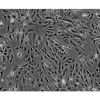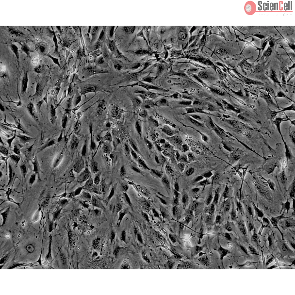
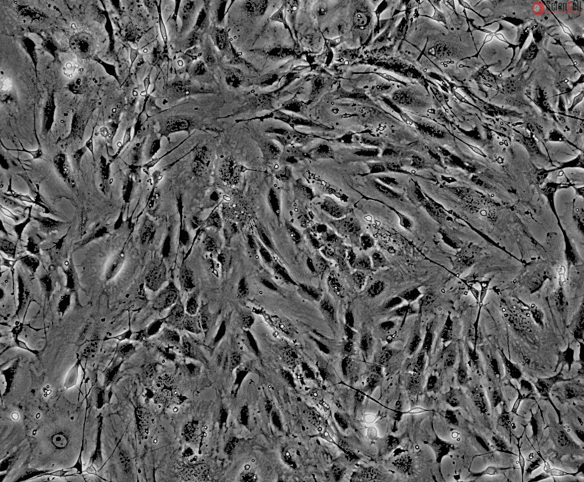
C57BL/6 小鼠来源的神经束膜成纤维细胞
该细胞分离自出生后第8天的 C57BL/6 小鼠神经组织。MPNF 在第1代(P1)进行冻存,并以冷冻形式交付。每支冻存管体积为 1 ml,细胞数量不少于 5 × 10⁵ 个。
Perineurial fibroblasts are of mesenchymal origin and form the perineurium. The perineurium plays an important role in maintaining the integrity of the internal peripheral nerve environment by creating a physical barrier that, under physiologic conditions, limits the entry of biologically active proteins, infectious agents, and blood-borne cells into the nerve bundles. The perineurial fibroblasts are characterized by distinct ultrastructural features, including non-branching thin cytoplasmic processes coated by an external lamina and joined at their ends by a tight junction, few organelles, actin and vimentin filaments, and numerous pinocytotic vesicles. Perineurial fibroblasts are initially recruited from the surrounding mesenchyme to form a loose, permeable sheath around axons and Schwann cells, where they are separated by the extracellular matrix. These cells later undergo a mesenchymal-to-epithelial transition to form tight junctions and organize into the perineurium. Perineurial fibroblasts are immunoreactive for vimentin and epithelial membrane antigen but not for the Schwann cell marker S-100.
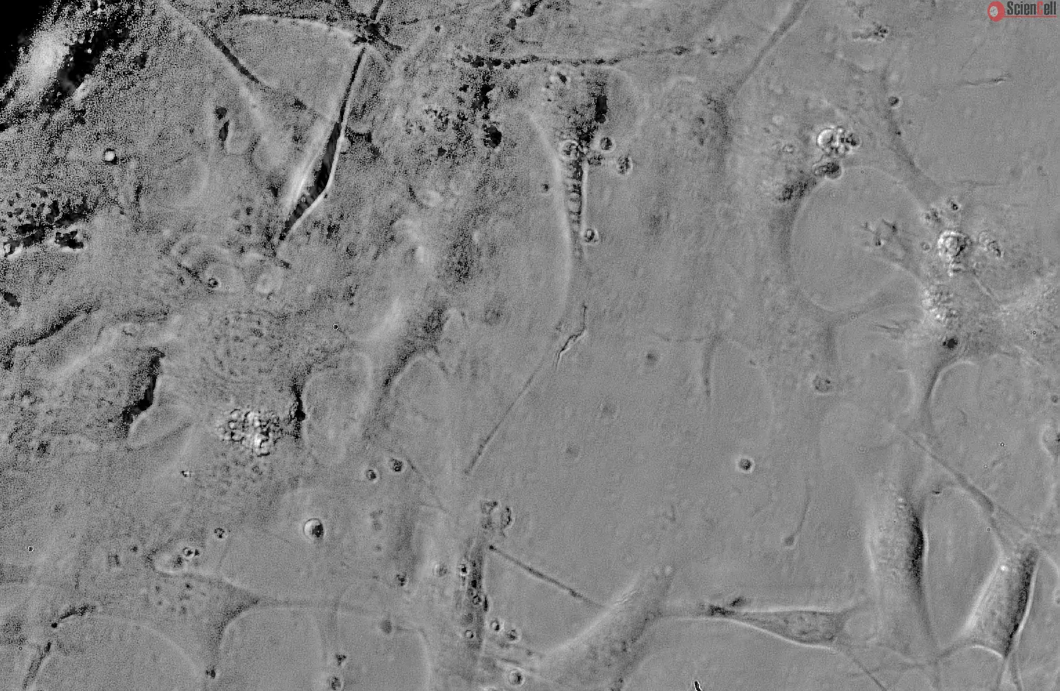
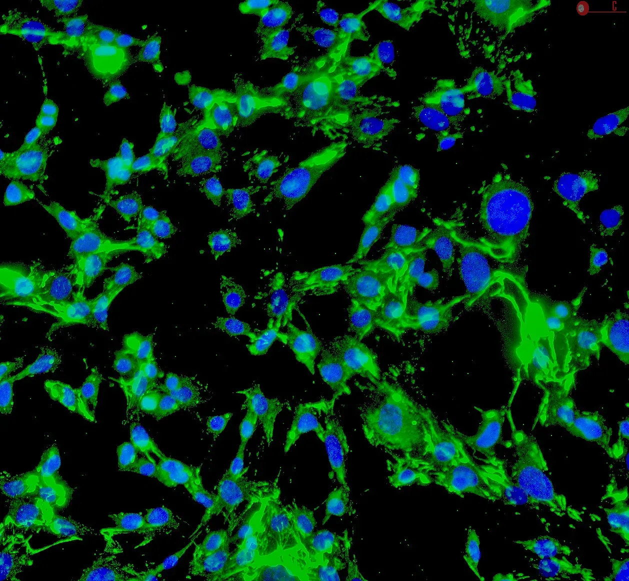
MPNF from ScienCell Research Laboratories are isolated from postnatal day 8 C57BL/6 mouse nerve. MPNF are cryopreserved at passage one and delivered frozen. Each vial contains >5 x 10⁵ cells in 1 ml volume. MPNF are characterized by immunofluorescence with antibodies specific to vimentin and/or fibronectin. MPNF are negative for mycoplasma, bacteria, yeast, and fungi. MPNF are guaranteed to further expand for 5 population doublings under the conditions provided by ScienCell Research Laboratories.
Recommended Medium
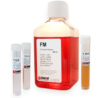
It is recommended to use Fibroblast Medium (FM, Cat. #2301) for culturing MPNF in vitro.
神经束膜成纤维细胞来源于间充质,并构成神经束膜。神经束膜通过形成一道物理屏障,在生理条件下限制生物活性蛋白、感染性因子以及血源性细胞进入神经束,从而在维持外周神经内部微环境的稳定性方面发挥关键作用。神经束膜成纤维细胞具有独特的超微结构特征,包括:不分支的细长细胞质突起,其表面覆盖外基膜,并在末端通过紧密连接相互连接;细胞器较少;含有肌动蛋白和波形蛋白丝;以及大量胞饮小泡。该类细胞最初由周围间充质募集而来,在轴突和施旺细胞周围形成一层松散且具有一定通透性的包裹结构,此时细胞之间由细胞外基质分隔。随后,这些细胞经历间充质向上皮样细胞的转化(mesenchymal-to-epithelial transition),逐渐形成紧密连接,并最终组织为结构完整的神经束膜。神经束膜成纤维细胞对波形蛋白(vimentin)和上皮膜抗原(epithelial membrane antigen)呈免疫反应阳性,而对施旺细胞标志物 S-100 呈阴性。
产品信息
ScienCell Research Laboratories 提供的小鼠神经束膜成纤维细胞(MPNF)分离自出生后第8天的 C57BL/6 小鼠神经组织。MPNF 在第一代传代时进行冻存,并以冻存形式运输。每瓶含有超过 5 × 10⁵ 个细胞,体积为 1 ml。MPNF 通过针对波形蛋白(vimentin)和/或纤维连接蛋白(fibronectin)的特异性抗体进行免疫荧光鉴定。该细胞产品经检测不含支原体、细菌、酵母和真菌。在 ScienCell 提供的培养条件下,MPNF 保证可继续扩增至少 5 个群体倍增。
推荐培养基
建议在体外培养 MPNF 时使用成纤维细胞培养基(Fibroblast Medium, FM,产品编号:2301)。
| 目录编号 | M1710-57 |
|---|---|
| 制造国家 | 美国 |
| 产品编码 | MPNF |
| 规格/数量 | 5 x 10^5 cells/vial |
| 产品用途 | 本产品仅供研究使用。未经批准,不得用于人体、动物或体外诊断程序。 |
| 储存 | 收到后,立即将细胞从干冰转移到液氮中,并在液氮中保存,直到需要进行实验。 |
| 运输 | 干冰。 |
Product Sheets

Search for a Certificate of Analysis by Lot Number below:
Find answers to common questions about ScienCell products, cell culture, handling, and research applications.
No Publication available at this time.