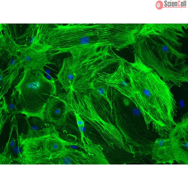
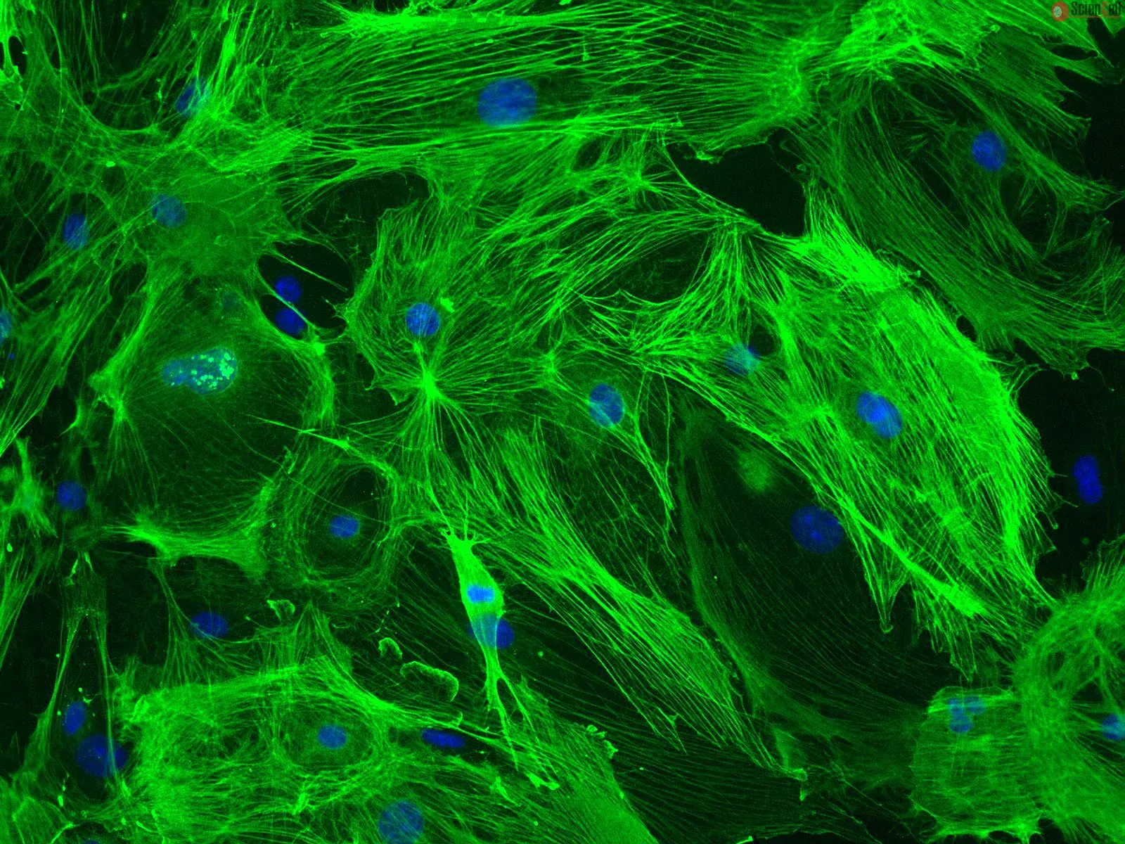
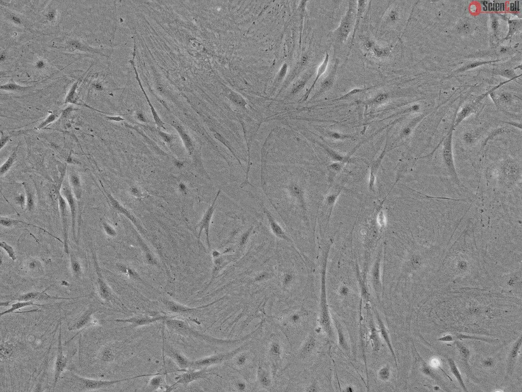
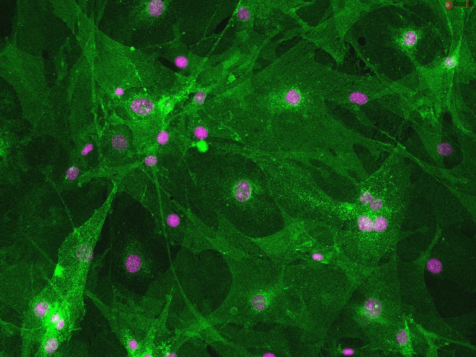
Brain Vascular Pericytes (BVP) are perivascular cells that are closely associated with the endothelium of capillaries and other small vessels. BVP, located between endothelial cells and astrocytes in the brain, communicate with other cells by extending long cytoplasmic processes which wrap around the capillaries. BVP participate in a variety of processes including angiogenesis, endothelial cell survival, regulation of capillary blood flow, and establishment and maintenance of the blood-brain barrier. Pericyte dysregulation has been linked to several pathological conditions such as hypertension, diabetic retinopathy, atherosclerosis, multiple sclerosis, Alzheimer’s disease, and tumor angiogenesis. The unique and diverse functions of BVP make them novel candidates for cell therapy in regenerative medicine. Cultured primary mouse BVP (MBVP) are a useful in vitro model for understanding the molecular mechanisms of blood-brain barrier regulation and for studying a wide variety of central nervous system diseases.
MBVP from ScienCell Research Laboratories are isolated from adult CD-1 mouse brain. MBVP are cryopreserved at passage one and delivered frozen. Each vial contains >5 x 10^5 cells in 1 ml volume. MBVP are characterized by immunofluorescence with antibody specific to plateletderived growth factor-β (PDGF Receptor-β) and α-smooth muscle actin. MBVP are negative for mycoplasma, bacteria, yeast, and fungi. MBVP are guaranteed to further expand for 5 population doublings under the conditions provided by ScienCell Research Laboratories.
Recommended Medium
It is recommended to use Pericyte Medium-mouse (PM, Cat. #1231) for culturing MBVP in vitro.
脑血管周细胞(BVP)是与毛细血管及其他小血管内皮细胞紧密相关的血管周围细胞。位于脑内皮细胞与星形胶质细胞之间的 BVP 可通过伸出长的细胞质突起包绕毛细血管,并与其他细胞进行通讯。BVP 参与多种生理过程,包括血管生成、内皮细胞存活、毛细血管血流调节以及血脑屏障的建立与维持。周细胞功能失调与多种病理状况相关,例如高血压、糖尿病视网膜病变、动脉粥样硬化、多发性硬化症、阿尔茨海默病以及肿瘤血管生成。BVP 所具有的独特且多样的功能,使其成为再生医学中细胞治疗的新型候选细胞。培养的原代小鼠 BVP(MBVP)是一种有价值的体外模型,可用于了解血脑屏障调控的分子机制,并研究多种中枢神经系统疾病。
ScienCell Research Laboratories 的小鼠脑血管周细胞(MBVP)分离自成年 CD-1 小鼠的脑组织。MBVP 在第 1 代时进行冻存,并以冷冻状态运输。每瓶含有超过 5 × 10^5 个细胞,体积为 1 ml。MBVP 采用针对血小板衍生生长因子-β(PDGF Receptor-β)和 α-平滑肌肌动蛋白的特异性抗体进行免疫荧光鉴定。MBVP 经检测不含支原体、细菌、酵母和真菌。在 ScienCell Research Laboratories 提供的培养条件下,MBVP 保证可进一步扩增 5 个群体倍增。
推荐培养基
建议使用 Pericyte Medium-mouse(PM,产品编号 #1231)在体外培养 MBVP。
Product Sheets
 M1200
Search for a Certificate of Analysis by Lot Number below:
Pericytes line the microvasculature throughout the body and play a key role in regulating blood flow by constricting and dilating vessels. However, the biophysical mechan... More
Pericytes line the microvasculature throughout the body and play a key role in regulating blood flow by constricting and dilating vessels. However, the biophysical mechanisms through which pericytes transduce microenvironmental chemical and mechanical cues to mediate vessel diameter, thereby impacting oxygen and nutrient delivery, remain largely unknown. This knowledge gap is clinically relevant as numerous diseases are associated with the aberrant contraction of pericytes, which are unusually susceptible to injury. Here, we report the development of a high-throughput hydrogel-based pericyte contraction cytometer that quantifies single-cell contraction forces from murine and human pericytes in different microvascular microenvironments and in the presence of competing vasoconstricting and vasodilating stimuli. We further show that murine pericyte survival in hypoxia is mediated by the mechanical microenvironment and that, paradoxically, pre-treating pericytes to reduce contraction increases hypoxic cell death. Moreover, using the contraction cytometer as a drug-screening tool, we found that cofilin-1 could be applied extracellularly to release murine pericytes from hypoxia-induced contractile rigor mortis and, therefore, may represent a novel approach for mitigating the long-lasting decrease in blood flow that occurs after hypoxic injury. Less
Chronic inflammation and blood–brain barrier dysfunction are key pathological hallmarks of neurological disorders such as multiple sclerosis, Alzheimer’s disease and ... More
Chronic inflammation and blood–brain barrier dysfunction are key pathological hallmarks of neurological disorders such as multiple sclerosis, Alzheimer’s disease and Parkinson’s disease. Major drivers of these pathologies include pro-inflammatory stimuli such as prostaglandins, which are produced in the central nervous system by the oxidation of arachidonic acid in a reaction catalyzed by the cyclooxygenases COX1 and COX2. Monoacylglycerol lipase hydrolyzes the endocannabinoid signaling lipid 2-arachidonyl glycerol, enhancing local pools of arachidonic acid in the brain and leading to cyclooxygenase-mediated prostaglandin production and neuroinflammation. Monoacylglycerol lipase inhibitors were recently shown to act as effective anti-inflammatory modulators, increasing 2-arachidonyl glycerol levels while reducing levels of arachidonic acid and prostaglandins, including PGE2 and PGD2. In this study, we characterized a novel, highly selective, potent and reversible monoacylglycerol lipase inhibitor (MAGLi 432) in a mouse model of lipopolysaccharide-induced blood–brain barrier permeability and in both human and mouse cells of the neurovascular unit: brain microvascular endothelial cells, pericytes and astrocytes. We confirmed the expression of monoacylglycerol lipase in specific neurovascular unit cells in vitro, with pericytes showing the highest expression level and activity. However, MAGLi 432 did not ameliorate lipopolysaccharide-induced blood–brain barrier permeability in vivo or reduce the production of pro-inflammatory cytokines in the brain. Our data confirm monoacylglycerol lipase expression in mouse and human cells of the neurovascular unit and provide the basis for further cell-specific analysis of MAGLi 432 in the context of blood–brain barrier dysfunction caused by inflammatory insults. Less
ScienCell Research Laboratories (SRL) takes pride in being a resource for researchers all over the world. The publications listed here are not meant as an endorsement or confirmation of the reliability of the products.