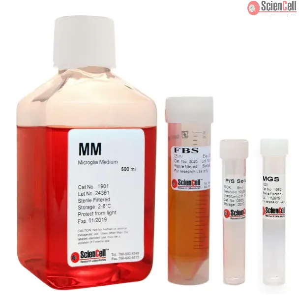
Microglia Medium (MM), when used with Microglia Growth Supplement (MGS, Cat #1952) and 25 ml of fetal bovine serum (FBS) is a complete medium designed for optimal growth of normal microglia in vitro. It is a sterile, liquid medium which contains essential and non-essential amino acids, vitamins, organic and inorganic compounds, hormones, growth factors, trace minerals and a low concentration of fetal bovine serum (5%). The medium is HEPES and bicarbonate buffered and has a pH of 7.4 when equilibrated in an incubator with an atmosphere of 5% CO2/95% air. The medium is formulated (quantitatively and qualitatively) to provide an optimally balanced nutritional environment that selectively supports the growth of normal microglia in vitro.
Endothelial cells lining the vessel network are indispensable for vascular transport but also provide paracrine signals controlling the behavior of nearby cell types. Per... More
Endothelial cells lining the vessel network are indispensable for vascular transport but also provide paracrine signals controlling the behavior of nearby cell types. Pericytes are another essential component of the vessel wall, but little is known about their interactions with other cell populations during organ growth and patterning. Here, we use mouse genetics to address the function of three pericyte-derived factors in postnatal lung and brain. We find that inactivation of the gene for hepatocyte growth factor (HGF) or brain-derived neurotrophic factor (BDNF) in pericytes causes no overt alterations in postnatal brain but impairs lung development, which we attribute to defective interaction with AT2 epithelial cells and pulmonary endothelium, respectively. In contrast, pericyte expression of the growth factor Nodal is dispensable for lung morphogenesis but regulates vessel growth and barrier function in the postnatal brain through interactions with endothelial cells, astrocytes and microglia. Taken together, our findings establish that pericytes are a critical source of paracrine signals controlling morphogenetic processes in an organ-specific fashion. Less
The lack of adequate human in vitro models that recapitulate the cellular composition and response of the human liver to injury hampers the development of anti-fibrotic d... More
The lack of adequate human in vitro models that recapitulate the cellular composition and response of the human liver to injury hampers the development of anti-fibrotic drugs. The goal of this study was to develop a human spheroid culture model to study liver fibrosis by using induced pluripotent stem cell (iPSC)-derived liver cells. iPSCs were independently differentiated towards hepatoblasts (iHepatoblasts), hepatic stellate cells (iHSCs), endothelial cells (iECs) and macrophages (iMΦ), before assembly into free floating spheroids by culturing cells in 96-well U-bottom plates and orbital shaking for up to 21 days to allow further maturation. Through transcriptome analysis, we show further maturation of iECs and iMΦ, the differentiation of the iHepatoblasts towards hepatocyte-like cells (iHeps) and the inactivation of the iHSCs by the end of the 3D culture. Moreover, these cultures display a similar expression of cell-specific marker genes (CYP3A4, PDGFRβ, CD31 and CD68) and sensitivity to hepatotoxicity as spheroids made using freshly isolated primary human liver cells. Furthermore, we show the functionality of the iHeps and the iHSCs by mimicking liver fibrosis through iHep-induced iHSC activation, using acetaminophen. In conclusion, we have established a reproducible human iPSC-derived liver culture model that can be used to mimic fibrosis in vitro as a replacement of primary human liver derived 3D models. The model can be used to investigate pathways involved in fibrosis development and to identify new targets for chronic liver disease therapy. Less
Microglia are the predominant resident central nervous system (CNS) cell type productively infected by HIV-1 and play a key role in the progression of HIV-associated deme... More
Microglia are the predominant resident central nervous system (CNS) cell type productively infected by HIV-1 and play a key role in the progression of HIV-associated dementia (HAD). Moreover, neural dysfunction and progression to HAD are accelerated in opiate drug abusers. In the present study, we examined the role of the autophagy pathway in the neuropathogenesis of HIV-1 using primary human microglial cells and determined whether opiates converge at this point. Infection of microglia with the HIV-1SF162 macrophage-tropic strain resulted in increased Beclin1 expression, accompanied by an increase in LC3 protein levels and accumulation of LC3 reporter RFP GFP (yellow) puncta, suggesting that HIV-1 infection triggers autophagosome formation without promoting protein degradation by the lysosome. Conversely, coexposure with HIV-1 and morphine significantly decreased virus-induced Beclin1 expression and autophagosome formation. Exploration of the possible mechanisms used by morphine to disrupt the autophagic process unveiled a significant increase in intracellular pH, which coincided with a reduction in the formation of acidic vesicular organelles and autophagolysosome formation. Small interfering RNA targeting BECN1, a gene critical for autophagosome formation, significantly reduced viral replication and the virus-induced inflammatory responses. Conversely, morphine-enhanced viral replication and inflammatory responses were not affected by gene silencing with siBeclin1, suggesting that the interactive effect of morphine in HIV-1 pathogenesis is mediated through a Beclin1-independent mechanism. These novel findings may have important implications for the connections between autophagy and HIV-1 pathogenesis mediated by microglial cells in opioid-abusing individuals. Less
ScienCell Research Laboratories (SRL) takes pride in being a resource for researchers all over the world. The publications listed here are not meant as an endorsement or confirmation of the reliability of the products.

 1901-b-prf (Basal & Phenol Red Free Medium)
,-25-ml.webp)