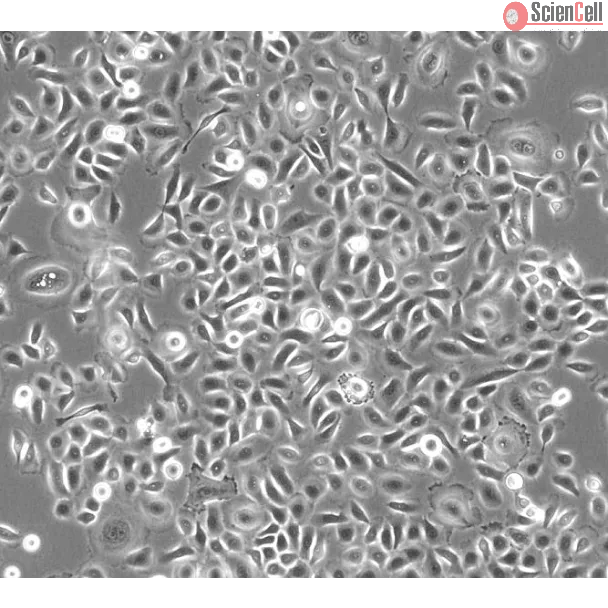
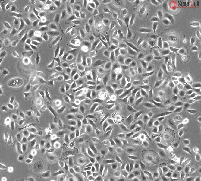
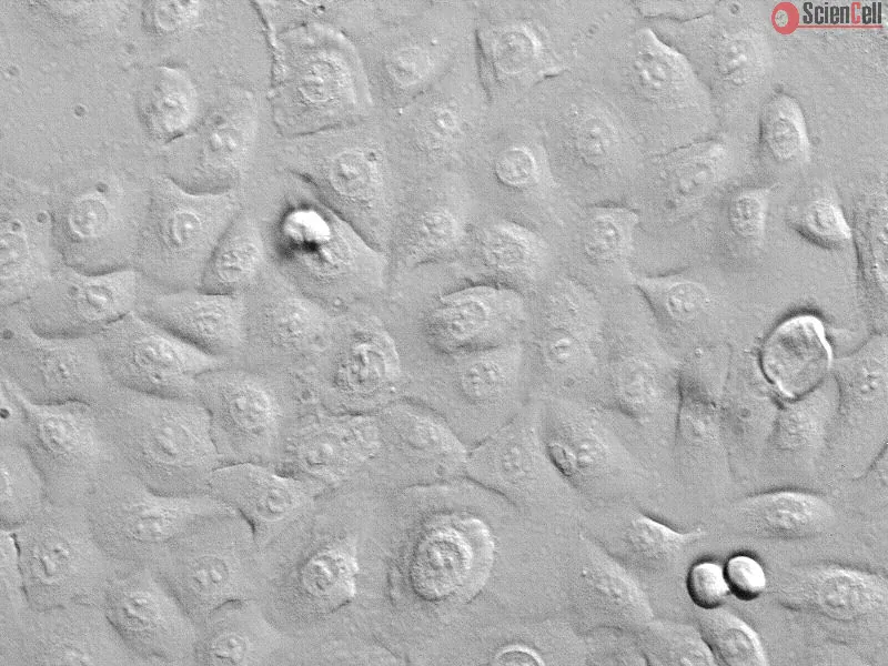
小气道位于肺泡与导气道之间的交界处。气道上皮细胞在气道中形成连续的衬里,作为外部有害物质的保护性物理和功能屏障,发挥着独特的作用。小气道上皮细胞(SAEpiC)通过产生趋化因子和表达黏附分子来调节免疫反应,帮助宿主防御。它们还产生液体,有助于肺部液体平衡。许多气道疾病,如哮喘、细支气管炎、慢性阻塞性肺病和囊性纤维化,都涉及气道表面上皮的损伤。研究人类SAEpiC可能有助于发现预防气道疾病的新治疗选择。
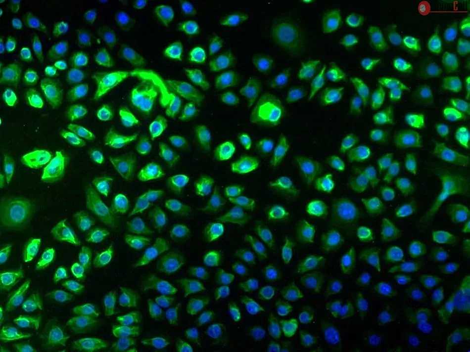
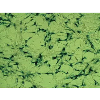
HPSAEpiC来自ScienCell研究实验室,分离自人类肺组织。HPSAEpiC在第一代时被冷冻保存并以冷冻状态交付。每个小瓶中含有>5 x 10⁵个细胞,体积为1毫升。HPSAEpiC通过免疫荧光染色表征,使用特异性抗体识别细胞角蛋白-18和/或细胞角蛋白-19。HPSAEpiC不含HIV-1、HBV、HCV、支原体、细菌、酵母和真菌。HPSAEpiC在ScienCell研究实验室提供的条件下保证可进一步扩增15倍人口倍增。
推荐培养基
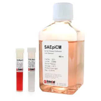
建议使用小气道上皮细胞培养基(SAEpiCM,Cat. #3231)来培养HPSAEpiC细胞。
Product Sheets
 3230
3230
Search for a Certificate of Analysis by Lot Number below:
The emergence of diseases associated with telomere dysfunction, including AIDS, aplastic anemia and pulmonary fibrosis, has bolstered interest in telomerase activators. W... More
The emergence of diseases associated with telomere dysfunction, including AIDS, aplastic anemia and pulmonary fibrosis, has bolstered interest in telomerase activators. We report identification of a new small molecule activator, GRN510, with activity ex vivo and in vivo. Using a novel mouse model, we tested the potential of GRN510 to limit fibrosis induced by bleomycin in mTERT heterozygous mice. Treatment with GRN510 at 10 mg/kg/day activated telomerase 2-4 fold both in hematopoietic progenitors ex vivo and in bone marrow and lung tissue in vivo, respectively. Telomerase activation was countered by co-treatment with Imetelstat (GRN163L), a potent telomerase inhibitor. In this model of bleomycin-induced fibrosis, treatment with GRN510 suppressed the development of fibrosis and accumulation of senescent cells in the lung via a mechanism dependent upon telomerase activation. Treatment of small airway epithelial cells (SAEC) or lung fibroblasts ex vivo with GRN510 revealed telomerase activating and replicative lifespan promoting effects only in the SAEC, suggesting that the mechanism accounting for the protective effects of GRN510 against induced lung fibrosis involves specific types of lung cells. Together, these results support the use of small molecule activators of telomerase in therapies to treat idiopathic pulmonary fibrosis. Less
Background: Epithelial-mesenchymal transition (EMT) plays a crucial role in small airway fibrosis of patients with chronic obstructive pulmonary disease (COPD). Increasin... More
Background: Epithelial-mesenchymal transition (EMT) plays a crucial role in small airway fibrosis of patients with chronic obstructive pulmonary disease (COPD). Increasing evidence suggests that the urokinase plasminogen activator receptor (uPAR) is involved in the pathogenesis of COPD. Increased uPAR expression has been implicated in the promotion of EMT in numerous cancers; however the role of uPAR in EMT in small airway epithelial cells of patients with COPD remains unclear. In this study, we investigated the degree of EMT and uPAR expression in lung epithelium of COPD patients, and verified the effect of uPAR on cigarette smoke extract (CSE)-induced EMT in vitro. Methods: The expression of EMT biomarkers and uPAR was assessed in lung epithelium specimens from non-smokers (n = 25), smokers (n = 25) and non-smokers with COPD (n = 10) and smokers with COPD (n = 18). The role of uPAR on CSE-induced EMT in human small airway epithelial cells (HSAEpiCs) was assessed by silencing uPAR expression in vitro. Results: Markers of active EMT and uPAR expression were significantly increased in the small airway epithelium of patients with COPD compared with controls. We also observed a significant correlation between uPAR and vimentin expression in the small airway epithelium. In vitro, CSE-induced EMT in HSAEpiCs was associated with high expression of uPAR, and targeted silencing of uPAR using shRNA inhibited CSE-induced EMT. Finally, we demonstrate that the PI3K/Akt signaling pathway is required for uPAR-mediated EMT in HSAEpiCs. Conclusions: A uPAR-dependent signaling pathway is required for CSE-induced EMT, which contributes to small airway fibrosis in COPD. We propose that increased uPAR expression in the small airway epithelium of patients with COPD participates in an active EMT process. Less
Background and purpose. Metal ion toxicity both locally and systemically following MoM hip replacements remains a concern. Cobalt ions have been shown to induce secretion... More
Background and purpose. Metal ion toxicity both locally and systemically following MoM hip replacements remains a concern. Cobalt ions have been shown to induce secretion of proinflammatory chemokines locally; however, little is known about their effect systemically. We investigated the in vitro effect of cobalt ions on a variety of cell lines by measuring production of the proinflammatory chemokines IL-8 and MCP-1. Less
ScienCell Research Laboratories (SRL) takes pride in being a resource for researchers all over the world. The publications listed here are not meant as an endorsement or confirmation of the reliability of the products.