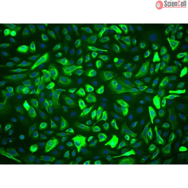
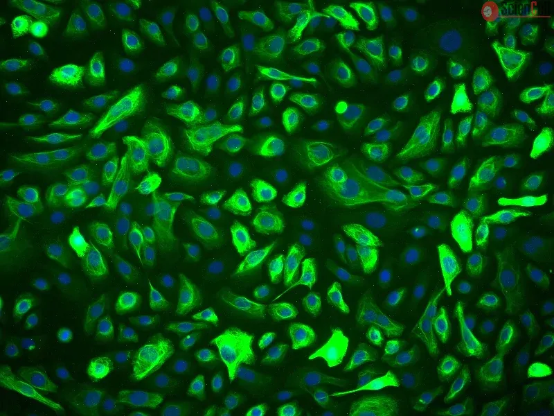
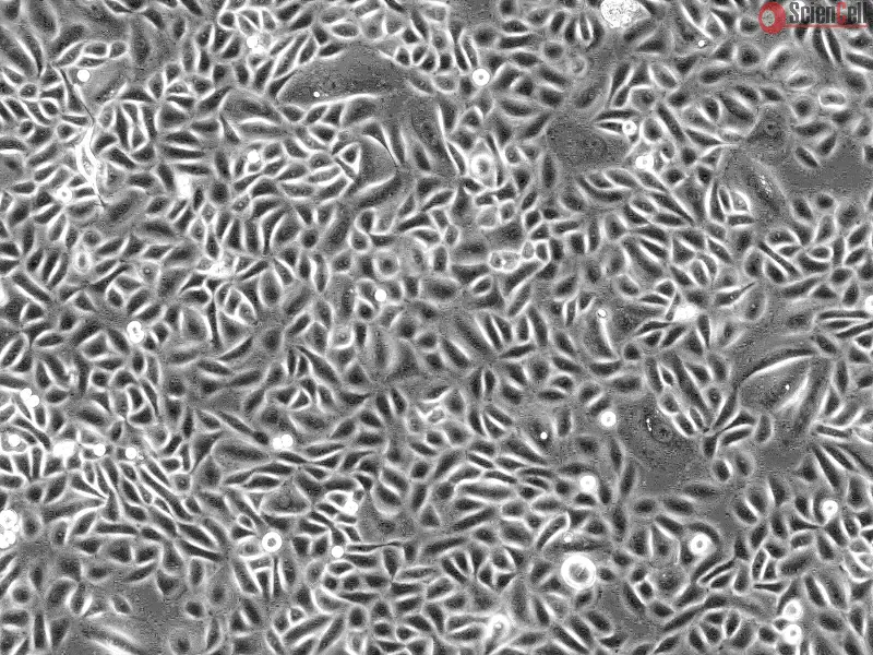
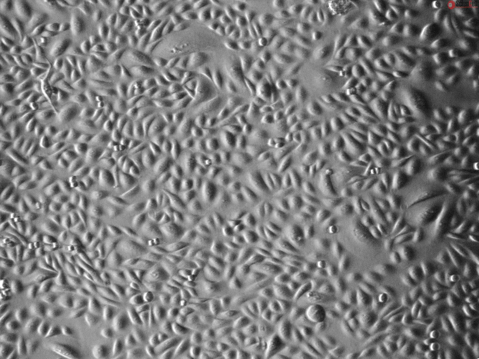
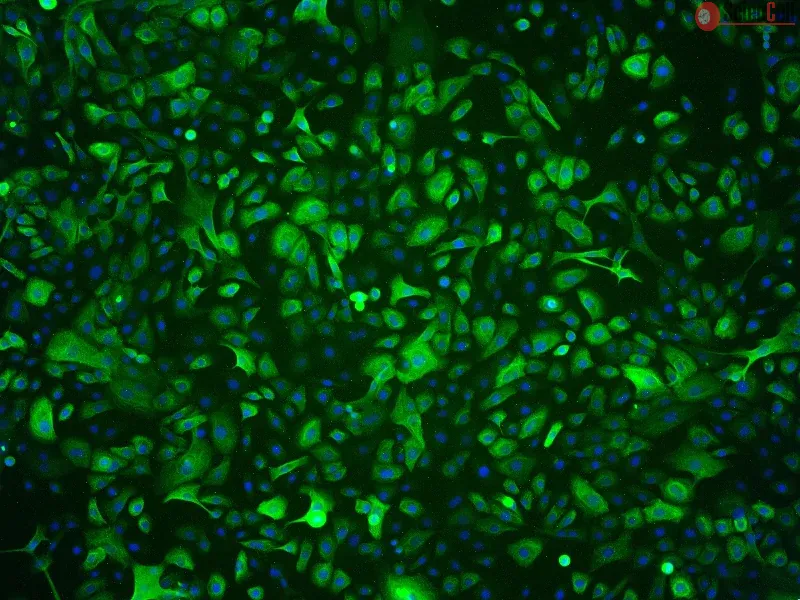
前列腺是雄性哺乳动物生殖系统中的主要附属器官,其结构和功能活动受雄激素控制。前列腺由三个区室组成,可通过细胞角蛋白表达谱加以区分。基底细胞主要表达高分子量细胞角蛋白(CK5 和 CK14),腺泡分泌细胞主要表达低分子量细胞角蛋白(CK8 和 CK18),而中间型细胞则同时表达基底和腺泡型细胞角蛋白。此外,越来越多的研究表明基底上皮细胞可以分化为腺泡上皮细胞。前列腺癌是影响男性最常见的癌症之一,其发病率和死亡率均较高。前列腺上皮细胞(PrEpiC)培养为研究前列腺的多项重要特性以及化学和激素致癌机制提供了独特的机会。
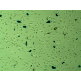
ScienCell Research Laboratories 的 HPrEpiC 是从正常人类前列腺组织中分离获得的。HPrEpiC 在第一代时经冻存保存,并以冷冻形式交付。每管含有 >5 x 10⁵ 个细胞,体积为 1 ml。HPrEpiC 经免疫荧光鉴定,使用特异性针对细胞角蛋白-18 和/或细胞角蛋白-19 的抗体。HPrEpiC 经检测阴性于 HIV-1、HBV、HCV、支原体、细菌、酵母菌和真菌。在 ScienCell 提供的培养条件下,HPrEpiC 保证可进一步扩增至 15 个人口倍增。
推荐培养基
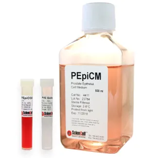
建议使用前列腺上皮细胞培养基(PEpiCM,货号 #4411)在体外培养 HPrEpiC。
Product Sheets
 4410
4410
Search for a Certificate of Analysis by Lot Number below:
Purpose
Bladder outlet obstruction (BOO) leads to detrusor overactivity (DO) and structural remodeling of the bladder. However, the molecular mechanisms underlying this ... More
Purpose
Bladder outlet obstruction (BOO) leads to detrusor overactivity (DO) and structural remodeling of the bladder. However, the molecular mechanisms underlying this process remain incompletely understood.
Materials and Methods
In a rat model of partial BOO, we assessed bladder function via cystometry and evaluated histological changes using hematoxylin and eosin staining. The expression and localization of aquaporin (AQP)-2 and caveolin (CAV)-1, -2, and -3 were analyzed by Western blotting and immunofluorescence in bladder tissues. Additionally, immunostaining was performed in cultured human bladder smooth muscle cells to assess protein co-localization.
Results
BOO rats exhibited elevated detrusor contraction pressure and shortened intercontraction intervals. Histologically, hypertrophied smooth muscle bundles and increased extracellular matrix were observed. Western blot analysis revealed significant upregulation of AQP2 and CAV1–3 in BOO bladders. Tissue immunofluorescence demonstrated increased expression and redistribution of these proteins in the detrusor muscle. Cultured cell analysis confirmed subcellular co-localization of AQP2 with CAV1–3.
Conclusions
DO induced by BOO is associated with upregulated expression of AQP2 and CAV1–3 in the urinary bladder. The coexpression observed in bladder cells suggests potential molecular interactions between AQP2 and CAVs that may contribute to the pathophysiology of BOO-related bladder dysfunction.
Keywords: Aquaporin-2; Bladder outlet obstruction; Caveolin; Urinary bladder, overactive Less
This study reports on probing the utility of in situ chromatin texture features such as nuclear DNA methylation and chromatin condensation patterns — visualized by fluo... More
This study reports on probing the utility of in situ chromatin texture features such as nuclear DNA methylation and chromatin condensation patterns — visualized by fluorescent staining and evaluated by dedicated three-dimensional (3D) quantitative and high-throughput cell-by-cell image analysis — in assessing the proliferative capacity, i.e. growth behavior of cells: to provide a more dynamic picture of a cell population with potential implications in basic science, cancer diagnostics/prognostics and therapeutic drug development. Two types of primary cells and four different cancer cell lines were propagated and subjected to cell-counting, flow cytometry, confocal imaging, and 3D image analysis at various points in culture. Additionally a subset of primary and cancer cells was accelerated into senescence by oxidative stress. DNA methylation and chromatin condensation levels decreased with declining doubling times when primary cells aged in culture with the lowest levels reached at the stage of proliferative senescence. In comparison, immortal cancer cells with constant but higher doubling times mostly displayed lower and constant levels of the two in situ-derived features. However, stress-induced senescent primary and cancer cells showed similar levels of these features compared with primary cells that had reached natural growth arrest. With regards to global DNA methylation and chromatin condensation levels, aggressively growing cancer cells seem to take an intermediate level between normally proliferating and senescent cells. Thus, normal cells apparently reach cancer-cell equivalent stages of the two parameters at some point in aging, which might challenge phenotypic distinction between these two types of cells. Companion high-resolution molecular profiling could provide information on possible underlying differences that would explain benign versus malign cell growth behaviors. Keywords: DNA methylation, chromatin condensation, cell proliferation, aging, senescence, cancer, 3D imaging, cell-by-cell analysis Less
ScienCell Research Laboratories (SRL) takes pride in being a resource for researchers all over the world. The publications listed here are not meant as an endorsement or confirmation of the reliability of the products.