Leydig Cells (LC) are located in the interstitium of the testis near the seminiferous tubules. One of the main functions of LC is to produce testosterone after stimulation by pituitary luteinizing hormone. LC, through synthesis of testosterone, play a key role in mammalian sex determination by inducing differentiation of the Wolffian ducts into male reproductive organs. Additionally, LC produce insulin-like growth factor 3 which is required for scrotal descent of the testis. Recent studies have indicated that molecules such as cyclic nucleotide phosphodiesterases can modulate testosterone production in LC, suggesting that testosterone synthesis may be manipulated using pharmacological targeting. Cultured Human LC are a useful in vitro model to better understand testicular development and to develop treatments for male reproductive disorders.
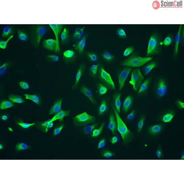
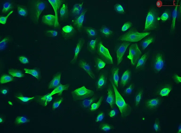
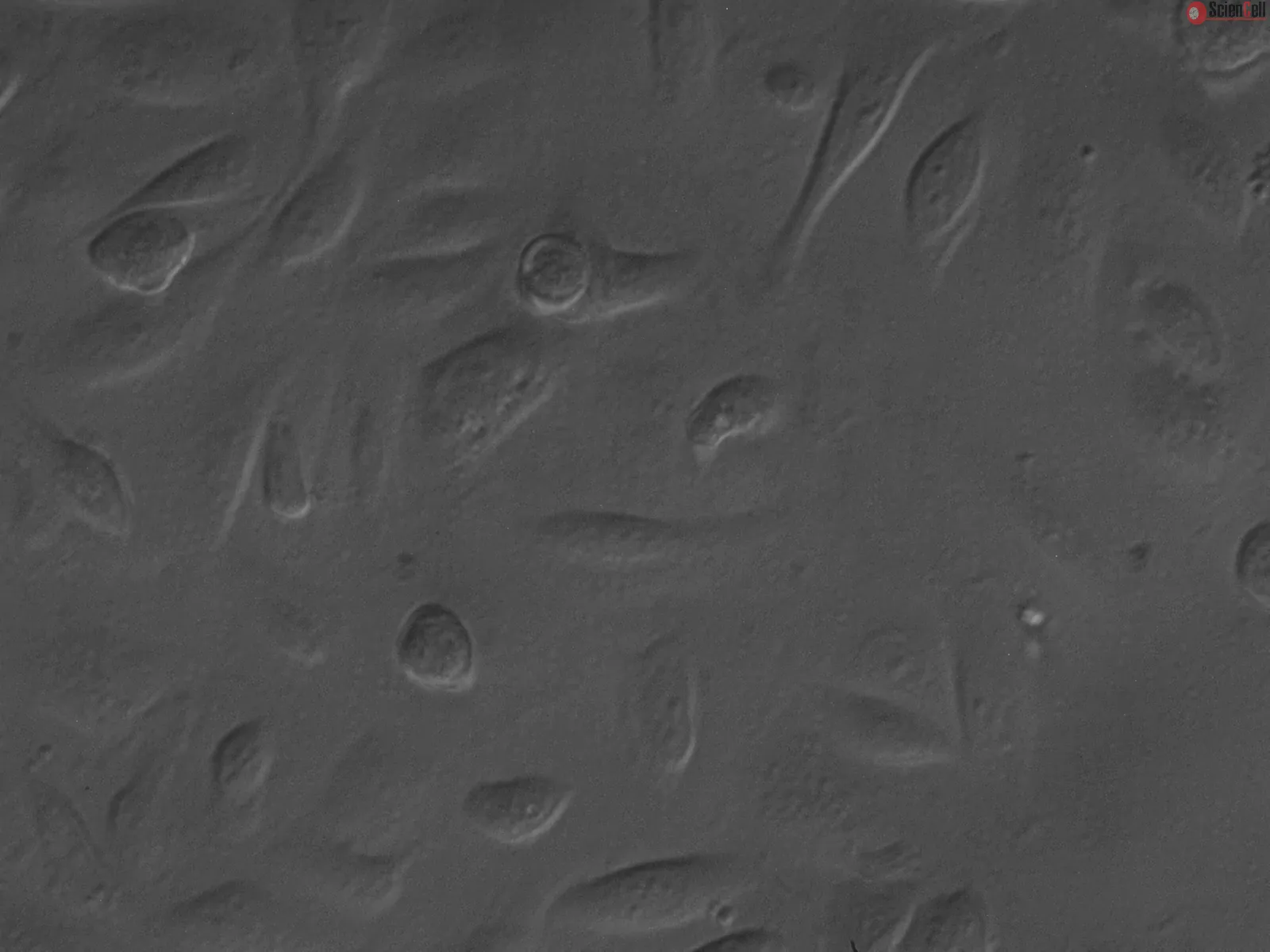
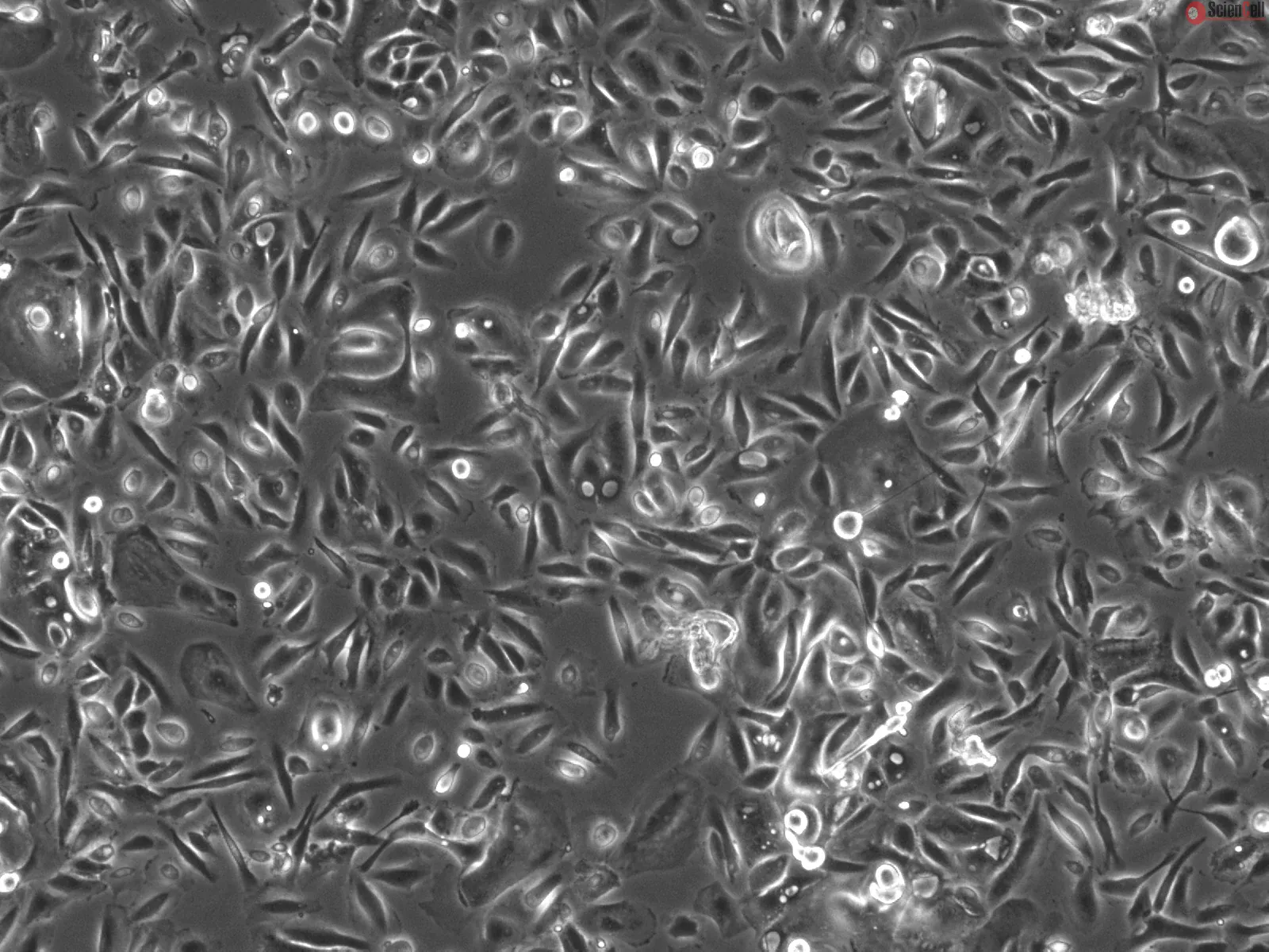
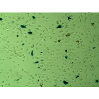
Human Leydig Cells (HLC) from ScienCell Research Laboratories are isolated from human testis. HLC are cryopreserved at passage one and delivered frozen. Each vial contains >5 x 10⁵ cells in 1 ml volume. HLC are characterized by immunofluorescence with antibody specific to GATA-4 and CK-18. HLC are negative for HIV-1, HBV, HCV, mycoplasma, bacteria, yeast and fungi. HLC are guaranteed to further culture under the conditions provided by ScienCell Research Laboratories; however, HLC are not recommended for expanding or long-term cultures due to limited expansion capacity.
Recommended Medium
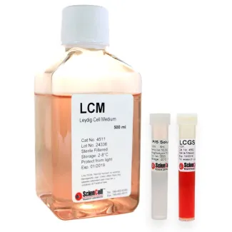
It is recommended to use Leydig Cell Medium (LCM, Cat. #4511) for the culturing of HLC in vitro.
睾丸间质细胞(Leydig 细胞,LC)位于睾丸间质中,邻近曲细精管。LC 的主要功能之一是在垂体黄体生成素(LH)的刺激下产生睾酮。LC 通过合成睾酮,在哺乳动物雄性性别分化过程中发挥重要作用,可促进沃尔夫管向男性生殖器官分化。此外,LC 还能产生胰岛素样生长因子 3(IGF3),该因子对于睾丸下降至阴囊至关重要。近期研究表明,环核苷酸磷酸二酯酶等分子可以调节 LC 的睾酮生成,提示可通过药理学靶向调控睾酮合成。培养的人睾丸间质细胞可作为有价值的体外模型,用于深入研究睾丸发育机制,并探索男性生殖系统疾病的治疗方法。
ScienCell Research Laboratories 的人睾丸间质细胞(HLC)分离自人睾丸组织。HLC 以第 1 代(P1)冻存,并以冷冻状态运输。每瓶含有 >5 × 10⁵ 个细胞,体积为 1 ml。HLC 采用针对 GATA-4 和 CK-18 的特异性抗体进行免疫荧光鉴定。HLC 经检测对 HIV-1、HBV、HCV、支原体、细菌、酵母菌和真菌均呈阴性。在 ScienCell Research Laboratories 提供的培养条件下,HLC 可进行进一步培养;但由于其扩增能力有限,不建议用于细胞扩增或长期培养。
推荐培养基
推荐使用睾丸间质细胞培养基(Leydig Cell Medium,LCM,产品编号:4511)在体外培养 HLC。
Product Sheets
 4510
Search for a Certificate of Analysis by Lot Number below:
The testicular interstitium relies on coordinated signaling among vascular, steroidogenic, and structural cells, yet the regulatory role of testicular endothelial cells (... More
The testicular interstitium relies on coordinated signaling among vascular, steroidogenic, and structural cells, yet the regulatory role of testicular endothelial cells (TECs) in maintaining this homeostasis remains unclear. Here, we identify TECs as a central signaling hub that orchestrates intercellular communication within the human testis. Integrative single-cell transcriptomic analysis of healthy and diabetic testes reveals that diabetes disrupts platelet-derived growth factor (PDGF) signaling. TECs in diabetes undergo endothelial-to-mesenchymal transition and exhibit reduced PDGFB expression, while Leydig and testicular peritubular cells downregulate PDGFRB, collectively weakening intercellular connectivity. This disruption silences the JUND-MCL1 survival program in Leydig cells, leading to apoptosis, extracellular matrix accumulation, and testosterone insufficiency, while impairing the contractility of testicular peritubular cells. Importantly, exogenous PDGF-BB supplementation reactivates the JUND-MCL1 axis, protects Leydig cells, alleviates fibrosis, and partially restores testosterone production and peritubular function. Together, these findings establish endothelial PDGF dysregulation as a key driver of diabetic testicular pathology and highlight PDGF-BB supplementation as a mechanistically grounded therapeutic strategy to restore interstitial and endocrine function in the context of diabetes. Less
Leydig cells are the primary source of testosterone or androgen production in male mammals. The blood–testis barrier (BTB) maintains structural integrity and safeguards... More
Leydig cells are the primary source of testosterone or androgen production in male mammals. The blood–testis barrier (BTB) maintains structural integrity and safeguards germ cells from harmful substances by blocking their entry into the seminiferous tubules. L-cysteine is essential to the production of glutathione, a powerful antioxidant crucial to protecting against oxidative stress-induced damage. Animal studies have demonstrated the protective effect of L-cysteine in preventing testicular damage caused by chemicals or radiation. This study examines whether L-cysteine enhances the expression of testosterone biosynthesis and the BTB genes in human Leydig cells and THP-1 monocytes. The Leydig cells and THP-1 monocytes were treated with L-cysteine for 24 h. RNA was extracted following treatment, and the gene expression was analyzed using quantitative RT-PCR. Testosterone levels in the cell supernatant were measured using an ELISA kit. L-cysteine treatment in Leydig cells significantly upregulated the expression of CYP11A1 (p = 0.03) and the BTB genes CLDN1 (p = 0.03), CLDN11 (p = 0.02), and TJP1 (p = 0.02). Similarly, L-cysteine significantly upregulated the expression of CYP11A1 (p = 0.03) and CYP19A1 (p < 0.01), and the BTB genes CLDN1 (p = 0.04), CLDN2 (p < 0.01), CLDN4 (p < 0.01), CLDN11 (p < 0.01), and TJP1 (p = 0.03) in THP-1 monocytes. Further, L-cysteine supplementation increased the testosterone secretion levels in human Leydig cells. The findings suggest that L-cysteine supplementation could be used as an adjuvant therapy to promote the integrity of the BTB genes, testosterone biosynthesis and secretion, and the maintenance of testicular functions, which in turn mitigates the risk of male infertility. Less
ScienCell Research Laboratories (SRL) takes pride in being a resource for researchers all over the world. The publications listed here are not meant as an endorsement or confirmation of the reliability of the products.