The keratocytes, or corneal fibroblasts, are highly specialized cells that are sandwiched between orthogonally arranged layers of collagen lamellae in the corneal stroma. They play a key role in maintaining the structure and transparency of the cornea, as they are the source of stromal collagen and proglycans. They also play important roles in corneal wound healing and tissue repair, and are known to undergo phenotypic transformations in wounds due to the influence of growth factors and cytokines. Under normal conditions, the keratocytes in the adult cornea are relatively quiescent cells. In the event of corneal injury or trauma, however, the keratocytes differentiate into active, synthesizing cells and rapidly replace damaged stromal matrix. Cultured human keratocytes express functional IL-4Rs and IL-17R on the cell surface, suggesting that these cells may contribute to the role of IL-4 and IL-17 as mediators of allergic reactions in the cornea. Changes in gene expression were observed in keratocytes after interleukin-1 treatment, which provides important insight into gene expression and suggests novel therapeutic targets for the control of corneal inflammation.
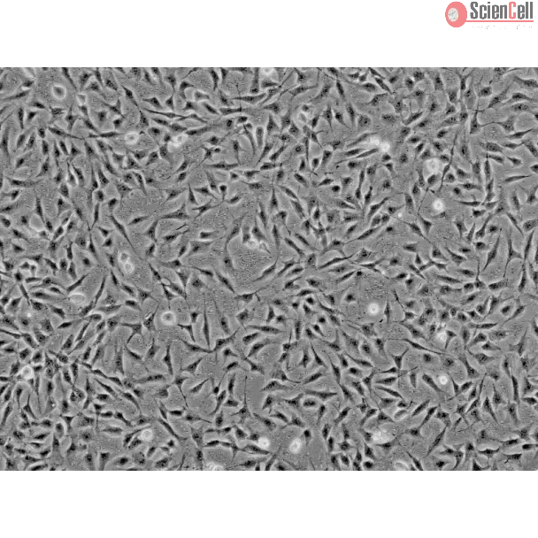
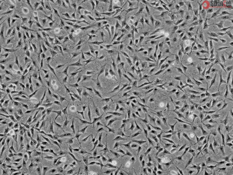
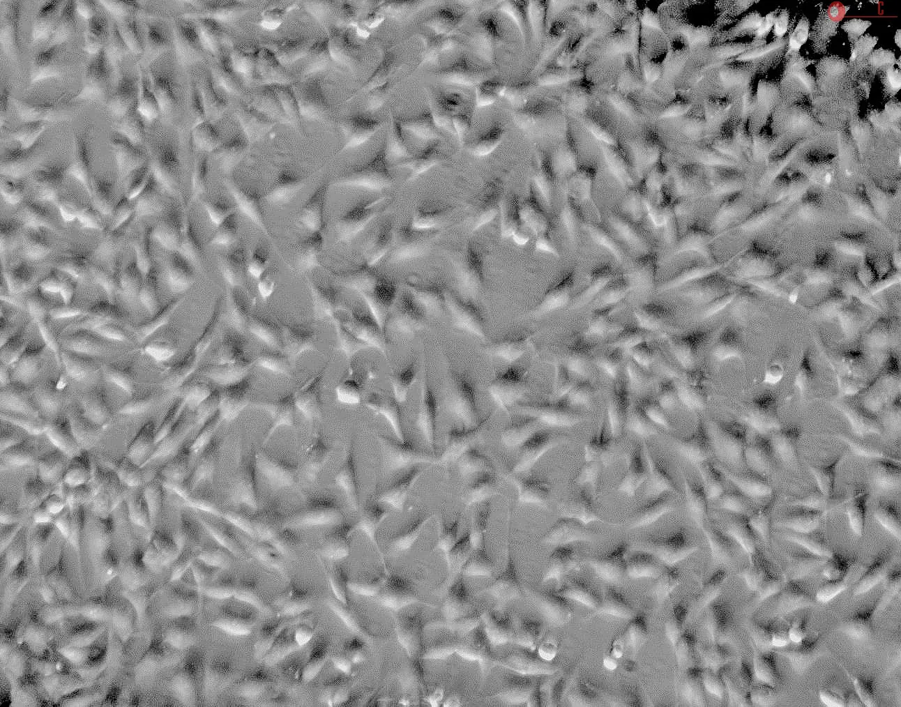
HK from ScienCell Research Laboratories are isolated from human cornea. HK are cryopreserved at passage one and delivered frozen. Each vial contains >5 x 10⁵ cells in 1 ml volume. HK are characterized by fibroblast morphology and immunofluorescence with antibody specific to fibronectin. HK are negative for HIV-1, HBV, HCV, mycoplasma, bacteria, yeast and fungi. HK are guaranteed to further expand for 15 population doublings under the conditions provided by ScienCell Research Laboratories.
Recommended Medium
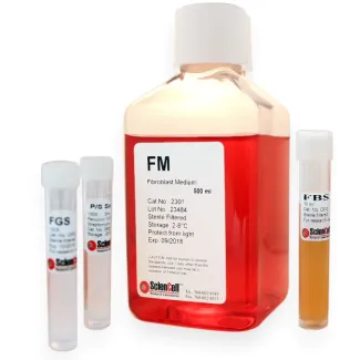
It is recommended to use Fibroblast Medium (FM, Cat. #2301) for culturing HK in vitro.
角膜基质细胞(keratocyte),又称角膜成纤维细胞,是高度特化的细胞,位于角膜基质中相互垂直排列的胶原板层之间。它们在维持角膜的结构和透明性方面发挥关键作用,是角膜基质胶原蛋白和蛋白聚糖的主要来源。此外,角膜基质细胞还参与角膜创伤愈合和组织修复,并可在生长因子和细胞因子的作用下,在损伤部位发生表型转变。正常情况下,成年角膜中的角膜基质细胞处于相对静息状态;但在角膜损伤或创伤发生后,这些细胞会分化为活化的合成细胞,并迅速补充受损的基质成分。培养的人角膜基质细胞表面表达具有功能性的 IL-4R 和 IL-17R,提示这些细胞可能参与介导 IL-4 和 IL-17 在角膜过敏反应中的作用。研究发现,白细胞介素-1(IL-1)处理后,角膜基质细胞的基因表达会发生变化,这为深入了解相关基因表达机制以及寻找控制角膜炎症的新型治疗靶点提供了重要依据。
ScienCell Research Laboratories 的 HK 分离自人角膜,于 P1 代进行冻存,并以冷冻状态提供。每瓶含有 >5 × 10⁵ 个细胞,体积为 1 mL。HK 通过成纤维细胞样形态以及采用针对纤连蛋白(fibronectin)的特异性抗体进行免疫荧光染色来表征。HK 经检测为 HIV-1、HBV、HCV、支原体、细菌、酵母菌和真菌阴性。在 ScienCell Research Laboratories 提供的培养条件下,HK 可进一步扩增至少 15 个群体倍增。
推荐培养基
建议使用成纤维细胞培养基(FM,产品编号 2301)进行 HK 的体外培养。
Product Sheets
 6520
6520
Search for a Certificate of Analysis by Lot Number below:
Corneal curvature abnormalities drive ectatic diseases, yet their mechanobiological effects on stromal cells remain poorly understood. We developed a hydraulically contro... More
Corneal curvature abnormalities drive ectatic diseases, yet their mechanobiological effects on stromal cells remain poorly understood. We developed a hydraulically controlled curvature array chip recapitulating disease-relevant geometries (33-56D) to investigate how keratocytes, fibroblasts, and myofibroblasts respond to geometric stress. Curvature-induced mechanical stress triggered dramatic cellular remodeling keratocytes exhibited significant proliferative enhancement and phenotypic transformation with ALDH3A1 downregulation and α-SMA upregulation, indicating mechanobiologically driven fibrotic activation. Fibroblasts developed curvature-dependent orthogonal alignment that recapitulates native corneal lamellar organization without chemical cues, while myofibroblasts showed enhanced contractile responses. RNA sequencing revealed that geometric stress activates identical molecular pathways dysregulated in keratoconus, including TGF-β/SMAD signaling, ECM-receptor interactions, and inflammatory cascades. Extracellular matrix remodeling was cell-type specific, with keratocytes showing homeostatic control loss, fibroblasts promoting matrix deposition, and myofibroblasts driving degradation. These findings establish curvature-induced mechanotransduction as the fundamental driver of corneal ectatic disease progression, repositioning geometric stress from a passive consequence to an active determinant of pathology. Less
Here a bioengineered platform is introduced to investigate adverse effects of environmental materials on the human cornea. Using primary cells, this system is capable of ... More
Here a bioengineered platform is introduced to investigate adverse effects of environmental materials on the human cornea. Using primary cells, this system is capable of reproducing the differentiated corneal epithelium and its underlying stroma in the human eye, which can then be treated with externally applied solid, liquid, or gaseous substances in a controlled manner and under physiologically relevant conditions. The proof-of-principle of how this system can be used to simulate human ocular exposure to different classes of environmental toxicants for direct visualization and quantitative analysis of their potential to induce acute corneal injury and inflammation is demonstrated. This model can also be further engineered to create an electromechanically actuated array of multiple human corneal tissues that can emulate spontaneous eye blinking. Using this advanced system, it is shown that blinking-like mechanical motions may play a protective role against adverse effects of environmental toxicants. This work yields an immediately deployable in vitro technology for screening ocular toxicity of existing and emerging environmental materials of various types and may enable the development of more realistic, human-relevant preclinical toxicology models complementary to traditional animal testing. Less
Keratoconus (KC) is a progressive degenerative disease that usually occurs bilaterally and is characterized by corneal thinning and apical protrusion of the cornea. Oxida... More
Keratoconus (KC) is a progressive degenerative disease that usually occurs bilaterally and is characterized by corneal thinning and apical protrusion of the cornea. Oxidative stress is an indicator of the accumulation of reactive oxygen species (ROS), and KC keratocytes exhibit increased ROS production compared with that of normal keratocytes. Therefore, oxidative stress in KC keratocytes may play a major role in the development and progression of KC. Here, we investigated the protective effect of sulforaphane (SF) antioxidants using a hydrogel-simulated model of the cell mechanical microenvironment of KC. The stiffness of the KC matrix microenvironment in vitro was 16.70 kPa and the stiffness of the normal matrix microenvironment was 34.88 kPa. Human keratocytes (HKs) were cultured for 24 h before observation or drug treatment with H2O2 in the presence or absence of SF. The levels of oxidative stress, nuclear factor E2-related factor 2 (Nrf-2) and antioxidant response element (ARE) were detected. The high-stress state of HKs in the mechanical microenvironment of KC cells compensates for the activation of the Nrf-2/ARE signaling pathway. H2O2 leads to increased oxidative stress and decreased levels of antioxidant proteins in KC. In summary, SF can reduce endogenous and exogenous oxidative stress and increase the antioxidant capacity of cells. Less
Background: Mutations in a novel gene, UBIAD1, were recently found to cause the autosomal dominant eye disease Schnyder corneal dystrophy (SCD). SCD is characterized by a... More
Background: Mutations in a novel gene, UBIAD1, were recently found to cause the autosomal dominant eye disease Schnyder corneal dystrophy (SCD). SCD is characterized by an abnormal deposition of cholesterol and phospholipids in the cornea resulting in progressive corneal opacification and visual loss. We characterized lesions in the UBIAD1 gene in new SCD families and examined protein homology, localization, and structure.Methodology/principal findings: We characterized five novel mutations in the UBIAD1 gene in ten SCD families, including a first SCD family of Native American ethnicity. Examination of protein homology revealed that SCD altered amino acids which were highly conserved across species. Cell lines were established from patients including keratocytes obtained after corneal transplant surgery and lymphoblastoid cell lines from Epstein-Barr virus immortalized peripheral blood mononuclear cells. These were used to determine the subcellular localization of mutant and wild type protein, and to examine cholesterol metabolite ratios. Immunohistochemistry using antibodies specific for UBIAD1 protein in keratocytes revealed that both wild type and N102S protein were localized sub-cellularly to mitochondria. Analysis of cholesterol metabolites in patient cell line extracts showed no significant alteration in the presence of mutant protein indicating a potentially novel function of the UBIAD1 protein in cholesterol biochemistry. Molecular modeling was used to develop a model of human UBIAD1 protein in a membrane and revealed potentially critical roles for amino acids mutated in SCD. Potential primary and secondary substrate binding sites were identified and docking simulations indicated likely substrates including prenyl and phenolic molecules.Conclusions/significance: Accumulating evidence from the SCD familial mutation spectrum, protein homology across species, and molecular modeling suggest that protein function is likely down-regulated by SCD mutations. Mitochondrial UBIAD1 protein appears to have a highly conserved function that, at least in humans, is involved in cholesterol metabolism in a novel manner. Less
Peroxisome proliferator-activated receptor α (PPARα) agonism in ocular inflammation has not been thoroughly investigated. The objective of this investigation was to det... More
Peroxisome proliferator-activated receptor α (PPARα) agonism in ocular inflammation has not been thoroughly investigated. The objective of this investigation was to determine the effect of WY-14 643, a selective PPARα agonist, on inflammatory cytokine release in human ocular cells. Stimulation of primary human corneal epithelial cells, keratocytes, and retinal endothelial cells with 1 to 10 ng/mL interleukin 1β (IL-1β) resulted in a significant increase in numerous inflammatory cytokines, including IL-6, IL-8, and tumor necrosis factor α (TNF-α); and dexamethasone was able to significantly inhibit these effects. However, WY-14 643 did not effectively block IL-1β-induced cytokine release in ocular cells; rather, significant increases in IL-1β-induced inflammatory cytokines were observed in these cells but not in aortic smooth muscle cells. WY-14 643 also significantly upregulated vascular endothelial growth factor (VEGF) expression in corneal epithelial cells and keratocytes. These studies demonstrate for the first time that PPARα agonism may be proinflammatory and proangiogenic in a variety of ocular cells and suggest that therapeutic applications of such agents in ophthalmology may be limited. Less
ScienCell Research Laboratories (SRL) takes pride in being a resource for researchers all over the world. The publications listed here are not meant as an endorsement or confirmation of the reliability of the products.