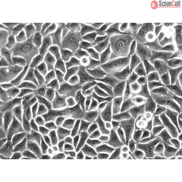
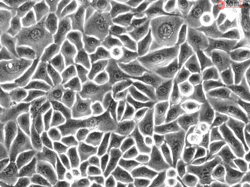
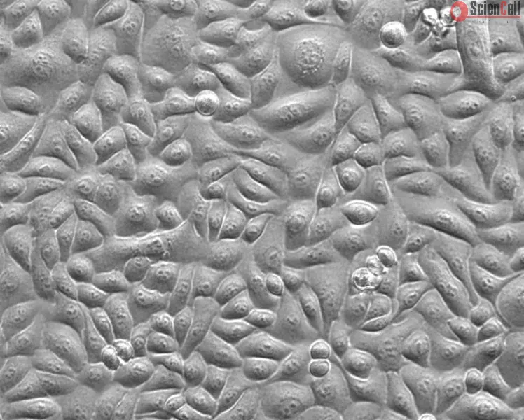
The human esophagus is lined by a non-keratinizing, stratified squamous epithelium whose apical cell membranes and intercellular junctional complexes form a barrier against the influx of luminal content. The barrier helps to reduce exposure of surface cells to changes in osmolality, which occurs frequently within the esophageal lumen. Histologically, the esophageal epithelium consists of two zones, the basal and differentiated zones. Cellular proliferation is limited to the basal zone, and the cells are thought to migrate from this area towards the esophageal lumen. Migration is associated with the initiation of differentiation and the sequential expression of differentiation markers. Human esophageal epithelial cells (HEsEpiC) are an excellent in vitro model to study esophageal epithelium physiology and the mechanisms of esophageal carcinogenesis.
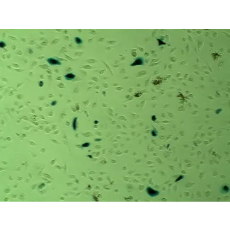
HEsEpiC from ScienCell Research Laboratories are isolated from the human esophagus. HEsEpiC are cryopreserved at passage one and delivered frozen. Each vial contains >5 x 10⁵ cells in 1 ml volume. HEsEpiC are characterized by immunofluorescence with antibody specific to cytokeratin-18 and/or cytokeratin-19. HEsEpiC are negative for HIV-1, HBV, HCV, mycoplasma, bacteria, yeast and fungi. HEsEpiC can be further cultured under the conditions provided by ScienCell Research Laboratories; however, HEsEpiC are not recommended for expanding or long-term cultures due to limited expansion capacity.
Recommended Medium
It is recommended to use Epithelial Cell Medium-2 (EpiCM-2, Cat. #4121) for culturing HEsEpiC in vitro.
人食管内壁由非角化复层鳞状上皮构成,其表层细胞膜及细胞间连接复合体形成一道屏障,可防止管腔内容物进入组织内部。该屏障有助于降低表层细胞受到渗透压变化的影响,而食管腔内经常会发生此类渗透压变化。从组织学角度来看,食管上皮由两个区域组成:基底层和分化层。细胞增殖主要发生于基底层,随后细胞被认为会从该区域向食管腔方向迁移。细胞迁移伴随着分化过程的启动以及分化标志物的依次表达。人食管上皮细胞(HEsEpiC)是研究食管上皮生理功能及食管癌发生机制的理想体外研究模型。
ScienCell Research Laboratories 提供的人食管上皮细胞(HEsEpiC)分离自人食管组织。HEsEpiC 在第一代(P1)阶段进行冻存,并以冷冻形式运输。每瓶含有超过 5 × 10⁵ 个细胞,总体积为 1 ml。HEsEpiC 通过使用针对细胞角蛋白-18(cytokeratin-18)和/或细胞角蛋白-19(cytokeratin-19)的特异性抗体进行免疫荧光染色鉴定。HEsEpiC 经检测不含 HIV-1、HBV、HCV、支原体、细菌、酵母菌及真菌。HEsEpiC 可在 ScienCell Research Laboratories 提供的培养条件下进一步培养;但由于其扩增能力有限,不建议用于大量扩增或长期培养。
推荐培养基
建议使用上皮细胞培养基-2(Epithelial Cell Medium-2,EpiCM-2,产品编号 #4121)对 HEsEpiC 进行体外培养。
Product Sheets
 2720
2720
Search for a Certificate of Analysis by Lot Number below:
Background YAP1, the nuclear effector of the Hippo pathway, has become an attractive target for treatment of malignancies and is a candidate oncogene in esophageal cancer... More
Background YAP1, the nuclear effector of the Hippo pathway, has become an attractive target for treatment of malignancies and is a candidate oncogene in esophageal cancer (EC). We hypothesized that knockdown of YAP1 could suppress EC and could be used for targeted therapy. However, there are few reports of the effect of YAP1 knockdown in EC. Materials and methods Quantitative real-time polymerase chain reaction and Western blot assays were performed to determine the expression levels of YAP1 mRNA and protein in primary EC tissue samples, EC cell lines, and controls. Immunohistochemistry was also performed to detect YAP1 protein expression in primary EC tumor and matched nontumor control tissues. YAP1-knockdown cell lines were constructed using short-hairpin RNA, and MTT, flow cytometry, and transwell chamber assays were used to analyze the effect of YAP1 knockdown on EC cell proliferation, apoptosis, and invasion. In vivo tumor formation assays were used to investigate the antitumor effect of YAP1 knockdown. Results We found that YAP1 mRNA and protein were upregulated in EC and that YAP1 expression correlated significantly with metastasis and tumor stage. We also found that YAP1 knockdown repressed cell proliferation and invasion and promoted apoptosis of EC cell lines. In addition, animal experiments revealed that YAP1 knockdown suppressed the growth of esophageal tumors in vivo. Conclusion Collectively, these data confirm our hypothesis that YAP1 knockdown suppresses EC and suggest that YAP1 knockdown could be exploited in the targeted gene therapy of EC in the future. Keywords: esophageal cancer, Hippo, YAP1 knockdown, animal experiments Less
Esophageal cancer is one of the most common malignant cancers worldwide. The molecular mechanism of esophageal squamous cell carcinoma (ESCC) is still poorly understood. ... More
Esophageal cancer is one of the most common malignant cancers worldwide. The molecular mechanism of esophageal squamous cell carcinoma (ESCC) is still poorly understood. ESE3 is a member of the Ets transcription family, which is only expressed in epithelial tissues and acts as a tumor suppressor gene in prostate cancer. Our study aim was to confirm whether ESE3 is involved in the carcinogenesis of ESCC. Immunohistochemical analysis revealed that ESE3 was mainly located in cell nuclei of normal tissues and the cytoplasm in ESCC tissues. Immunofluorescence and western blot analyses of the normal esophageal cell line HEEpiC and ESCC cell lines EC9706 TE-1, KYSE150, and KYSE410 confirmed these results. pEGFP-ESE3 and pcDNA3.1-V5/HisA-ESE3 plasmids were constructed for overexpression of ESE3 in EC9706 and KYSE150 cells. The stably transfected cells showed restoration of the nuclear localization of ESE3. EC9706 cells with re-localization of ESE3 to the nucleus showed inhibition of proliferation, colony formation, migration, and invasion. To explore the possible mechanism of the differences in localization of ESE3 in normal esophageal cells and ESCC cells, ESCC cell lines were treated with the nuclear export inhibitor leptomycin B, transcription inhibitor actinomycin D, PKC inhibitor sphinganine, P38 MAPK inhibitor SB202190, and CK II inhibitor TBCA. These reagents were chosen according to the well-known mechanisms of protein translocation. However, the localization of ESE3 was unchanged after these treatments. The sequence of ESE3 cDNA in ESCC cells was identical to the standard sequence of ESE3 in the NCBI Genebank database, indicating that there was no mutation in the coding region of ESE3 in ESCC. Taken together, our study suggests that ESE3 plays an important role in the carcinogenesis of ESCC through changes in subcellular localization and may act as a tumor suppressor gene in ESCC, although the mechanisms require further study. Less
ScienCell Research Laboratories (SRL) takes pride in being a resource for researchers all over the world. The publications listed here are not meant as an endorsement or confirmation of the reliability of the products.