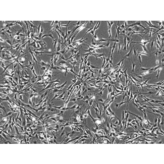
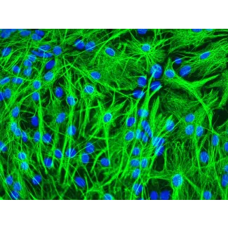
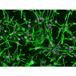
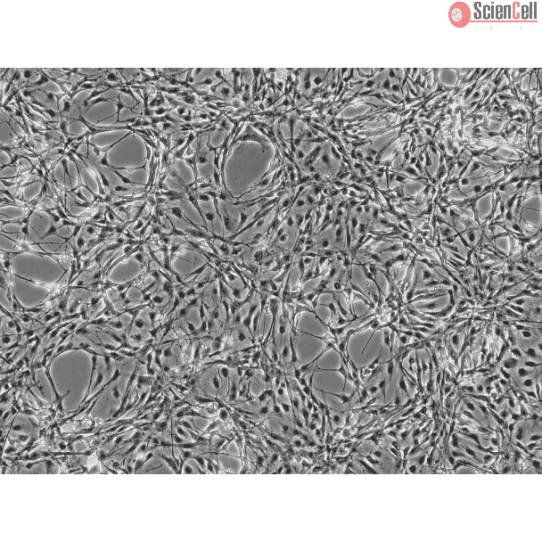
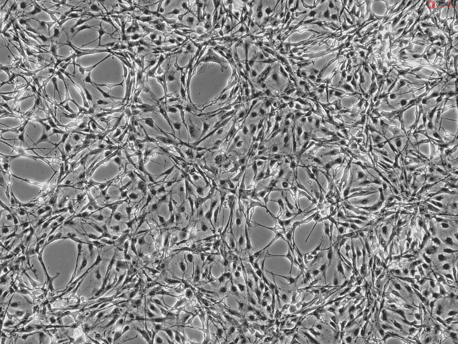
Astrocytes are the major cell type in the mammalian brain. They provide a variety of supportive functions to their partner neurons in the central nervous system (CNS), such as neuronal guidance during development, nutritional and metabolic support throughout life. Astrocytes have also been implicated in various pathological processes. Impairment of normal astrocyte functions during stroke and other insults can critically influence neuron survival. Long-term recovery after brain injury, through neurite outgrowth, synaptic plasticity, or neuron regeneration, is also influenced by astrocyte surface molecule expression and trophic factor release. Numerous studies have demonstrated that astrocytes are among the most functionally diverse group of cells in the CNS. Much of what we have learned about astrocytes is from in vitro studies and astrocyte culture is a useful tool for exploring the diverse properties of this cell type.
RA from ScienCell Research Laboratories are isolated from CD® IGS rat brain. RA are cryopreserved as primary cultures and delivered frozen. Each vial contains >5 x 10⁵ cells in 1 ml volume. RA are characterized by immunofluorescence with antibody specific to GFAP. RA are negative for mycoplasma, bacteria, yeast, and fungi. RA are guaranteed to further expand for 5 population doublings under the conditions provided by ScienCell Research Laboratories.
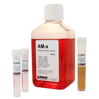
Recommended Medium
It is recommended to use Astrocyte Medium-animal (AM-a, Cat. #1831) for culturing RA in vitro.
Traumatic brain injury (TBI) is a major cause of morbidity and mortality worldwide, affecting over 10 million people annually, with an estimated cost of $76.5 billion. Al... More
Traumatic brain injury (TBI) is a major cause of morbidity and mortality worldwide, affecting over 10 million people annually, with an estimated cost of $76.5 billion. Although apocynin freely transverses the blood–brain barrier (BBB), its application is limited due to its rapid elimination, low terminal half-life (t1/2 = 6.7 min), narrow dose–response relationship, and cytotoxicity, thereby requiring repeated dosages. With this study, we aimed to develop transferrin-functionalized nanoparticles encapsulating apocynin to treat neuroinflammation for targeted drug delivery to sites of brain injury. As a preliminary approach, we endeavored to optimize the formulation parameters of apocynin-loaded albumin nanoparticles prepared through the desolvation method. The nanoparticles were characterized for their size, polydispersity, surface charge, drug loading and in vitro drug release. In this study, we also investigated the anti-inflammatory and neuroprotective effects of free apocynin and nanoparticle-loaded apocynin in neuronal cells. We show that the developed formulation displayed monodispersed, nanosized particles with higher entrapment efficiency, loading, stability, and sustained release profiles. The permeability of the nanoparticles across HBMECs reached the maximum at 67%. The in vivo evaluation revealed the enhanced uptake of transferrin-anchored nanoparticles in the brain tissues when compared with unmodified nanoparticles after I.V. administration. In vivo nanoparticle localization studies using a blast TBI (bTBI) model and confocal fluorescence microscopy have shown that tf-apoANPs are successful in delivering relatively high amounts of nanoparticles to the brain parenchyma and glial cells compared to non-targeted nanoparticles. We also establish that targeted nanoparticles accumulate in the brain. In conclusion, tf-apoANPs are efficacious carriers for targeted delivery across the blood–brain barrier to potentially treat neuroinflammation in brain injury and other diseases.
Keywords:
apocynin; nanoparticle; albumin; HPLC; desolvation method; targeted delivery; biodistribution; neuroprotection Less
Injury to the vertebrate central nervous system (CNS) induces astrocytes to change their morphology, to increase their rate of proliferation, and to display directional m... More
Injury to the vertebrate central nervous system (CNS) induces astrocytes to change their morphology, to increase their rate of proliferation, and to display directional migration to the injury site, all to facilitate repair. These astrocytic responses to injury occur in a clear temporal sequence and, by their intensity and duration, can have both beneficial and detrimental effects on the repair of damaged CNS tissue. Studies on highly regenerative tissues in non-mammalian vertebrates have demonstrated that the intensity of direct-current extracellular electric fields (EFs) at the injury site, which are 50–100 fold greater than in uninjured tissue, represent a potent signal to drive tissue repair. In contrast, a 10-fold EF increase has been measured in many injured mammalian tissues where limited regeneration occurs. As the astrocytic response to CNS injury is crucial to the reparative outcome, we exposed purified rat cortical astrocytes to EF intensities associated with intact and injured mammalian tissues, as well as to those EF intensities measured in regenerating non-mammalian vertebrate tissues, to determine whether EFs may contribute to the astrocytic injury response. Astrocytes exposed to EF intensities associated with uninjured tissue showed little change in their cellular behavior. However, astrocytes exposed to EF intensities associated with injured tissue showed a dramatic increase in migration and proliferation. At EF intensities associated with regenerating non-mammalian vertebrate tissues, these cellular responses were even more robust and included morphological changes consistent with a regenerative phenotype. These findings suggest that endogenous EFs may be a crucial signal for regulating the astrocytic response to injury and that their manipulation may be a novel target for facilitating CNS repair. Less
ScienCell Research Laboratories (SRL) takes pride in being a resource for researchers all over the world. The publications listed here are not meant as an endorsement or confirmation of the reliability of the products.


,-1-mg-ml--2.jpg)