The coronary arteries provide a substantial blood supply to meet the enormous energy demands of the heart for its constant contractile activity. Lining the vessel wall, coronary artery endothelial cells (CAEC) are continuously exposed to fluid shear stress that induces changes in cell morphology and the production of endothelium-derived substances regulating vasoconstriction and vessel growth. CAEC also modulate the expression of cellular adhesion molecules to control and fine-tune inflammatory responses and fibrinolysis. These physiological properties allow CAEC cultures to be widely used in the study of mechanisms for endothelium dysfunction, pathogenesis of coronary heart disease and atherosclerosis, and the development of novel disease treatments.
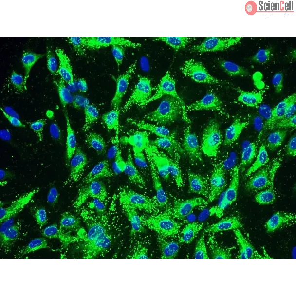
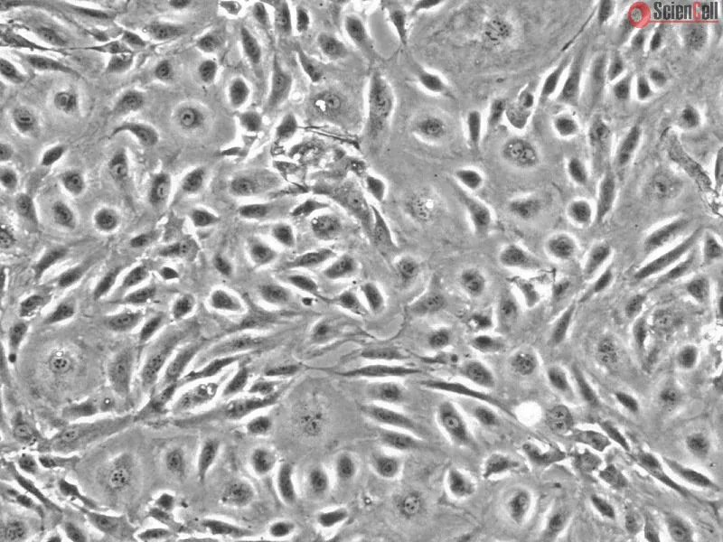
HCAEC from ScienCell Research Laboratories are isolated from human coronary artery. HCAEC are cryopreserved at passage one and delivered frozen. Each vial contains >5 x 10⁵ cells in 1 ml volume. HCAEC are characterized by immunofluorescence with antibodies specific to vWF/Factor VIII and/or CD31 (PECAM1). HCAEC are negative for HIV-1, HBV, HCV, mycoplasma, bacteria, yeast, and fungi. HCAEC are guaranteed to further expand for 10 population doublings under the conditions provided by ScienCell Research Laboratories.
Recommended Medium
It is recommended to use Endothelial Cell Medium (ECM, Cat. #1001) for culturing HCAEC in vitro.
High density lipoprotein (HDL) has been proposed to be internalized and to promote reverse cholesterol transport in endothelial cells (ECs). However, the mechanism underl... More
High density lipoprotein (HDL) has been proposed to be internalized and to promote reverse cholesterol transport in endothelial cells (ECs). However, the mechanism underlying these processes has not been studied. In this study, we aim to characterize HDL internalization and cholesterol efflux in ECs and regulatory mechanisms. We found mature HDL particles were reduced in patients with coronary artery disease (CAD), which was associated with an increase in CC-chemokine ligand 2 (CCL2). In cultured primary human coronary artery endothelial cells and human umbilical vein endothelial cells, we determined that CCL2 suppressed the binding (4 °C) and association (37 °C) of HDL to/with ECs and HDL cellular internalization. Furthermore, CCL2 inhibited [3H]cholesterol efflux to HDL/apoA1 in ECs. We further found that CCL2 induced CC-chemokine receptor 2 (CCR2) expression and siRNA-CCR2 reversed CCL2 suppression on HDL binding, association, internalization, and on cholesterol efflux in ECs. Moreover, CCL2 induced p42/44 mitogen-activated protein kinase (MAPK) phosphorylation via CCR2, and p42/44 MAPK inhibition reversed the suppression of CCL2 on HDL metabolism in ECs. Our study suggests that CCL2 was elevated in CAD patients. CCL2 suppressed HDL internalization and cholesterol efflux via CCR2 induction and p42/44 MAPK activation in ECs. CCL2 induction may contribute to impair HDL function and form atherosclerosis in CAD.
Keywords
atherosclerosis, cholesterol metabolism, endothelial cell, high-density lipoprotein (HDL), mitogen-activated protein kinase (MAPK), CC-chemokine ligand 2 (CCL2) Less
The present study investigated whether atorvastatin antagonizes the visfatin-induced expression of inflammatory mediators in human coronary artery endothelial cells (HCAE... More
The present study investigated whether atorvastatin antagonizes the visfatin-induced expression of inflammatory mediators in human coronary artery endothelial cells (HCAECs). Several analysis methods, such as reverse transcription-quantitative polymerase chain reaction, western blot analysis and H2DCFDA incubation, were used in the present study. The data showed that atorvastatin decreased the visfatin-induced expression of interleukin (IL)-6 and IL-8 in HCAECs. In addition, atorvastatin inhibited the visfatin-induced expression of intercellular adhesion molecule-1 and vascular cell adhesion molecule-1 in HCAECs. In addition, the present study found that atorvastatin inhibited the visfatin-activated nuclear factor-κB (NF-κB) signal pathway by preventing extracellular signal-regulated kinase phosphorylation in HCAECs. Atorvastatin significantly inhibited visfatin-induced NF-κB activity via the upregulation of reactive oxygen species production. Atorvastatin, a visfatin antagonist (FK866) and an NF-κB inhibitor (BAY11-7082) decreased the visfatin-induced expression of inflammatory mediators via the upregulation of NF-κB activation in HCAECs. These results suggest that atorvastatin may inhibit
the visfatin‑induced upregulation of inflammatory mediators through blocking the NF-κB signal pathway. The findings of the present study provide a potential use for atorvastatin and visfatin in the pathogenesis of HCAEC dysfunction. This knowledge may contribute to the development of novel therapies for atherosclerosis. Less
The aim of this study was to investigate and characterize the efficacy and mechanism of action of asiaticoside in combination with rapamycin in the inhibition of in-stent... More
The aim of this study was to investigate and characterize the efficacy and mechanism of action of asiaticoside in combination with rapamycin in the inhibition of in-stent restenosis (ISR). The effects of asiaticoside combined with rapamycin on cell proliferation in vitro were evaluated by MTT assay. The mRNA expression was analyzed by quantitative polymerase chain reaction (qPCR). Enzyme-linked immunosorbent assay (ELISA) was used to confirm protein synthesis. The cell growth inhibition rate in the combination group was significantly higher compared with those in the asiaticoside and rapamycin groups for human aortic fibroblasts (HAFs; 63.50±3.83, 53.06±8.10 and 60.34±4.9%, respectively) and human aortic smooth muscle cells (HASMCs; 33.12±1.35, 26.21±7.59 and 28.27±4.92, respectively; P<0.05). However, for human coronary artery endothelial cells (HCAECs), the cell growth inhibition rates in the combination, asiaticoside and rapamycin groups were 11.09±1.17, 26.22±4.24 and 34.80±2.80%, respectively (P<0.05), as detected by MTT assay. The qPCR assay showed that in the combination group the level of von Willebrand factor (vWF) mRNA was downregulated, while platelet endothelial cell adhesion molecule (PECAM-1) and endothelial nitric oxide synthase (eNOS) mRNAs were upregulated in HCAECs compared with the rapamycin group (P<0.05). Transforming growth factor (TGF)-β1 and TIMP1 mRNAs were downregulated while Smad7 and matrix metalloproteinase 1 (MMP1) mRNAs were upregulated in HAFs compared with the rapamycin and AT groups (P<0.05). The ELISA showed that the type I collagen level was significantly reduced in HASMCs and HAFs (P<0.05). The data suggest that asiaticoside combined with rapamycin may be effective in the reduction of ISR. Keywords: in-stent restenosis, asiaticoside, rapamycin, Smad7 Less
ScienCell Research Laboratories (SRL) takes pride in being a resource for researchers all over the world. The publications listed here are not meant as an endorsement or confirmation of the reliability of the products.