Astrocytes are the major cell type in the mammalian brain. They provide a variety of supportive functions to their partner neurons in the central nervous system (CNS), such as neuronal guidance during development, nutritional and metabolic support throughout life. Astrocytes have also been implicated in various pathological processes. Impairment of normal astrocyte functions during stroke and other insults can critically influence neuron survival. Long-term recovery after brain injury, through neurite outgrowth, synaptic plasticity, or neuron regeneration, is also influenced by astrocyte surface molecule expression and trophic factor release. Numerous studies have demonstrated that astrocytes are among the most functionally diverse group of cells in the CNS. Much of what we have learned about astrocytes is from in vitro studies and astrocyte culture is a useful tool for exploring the diverse properties of this cell type.
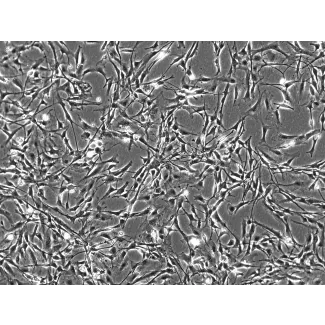
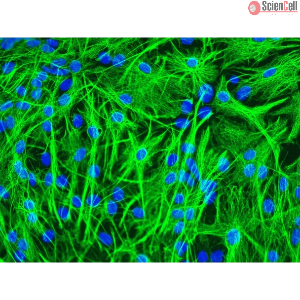
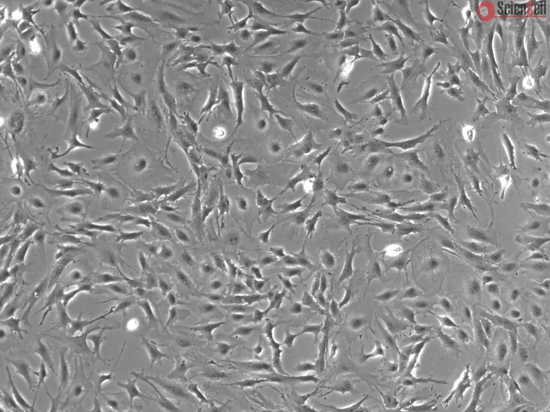
MA from ScienCell Research Laboratories are isolated from C57BL/6 mouse brain. MA are cryopreserved at P0 and delivered frozen. Each vial contains >5 x 10⁵ cells in 1 ml volume. MA are characterized by immunofluorescence with antibody specific to GFAP. MA are negative for mycoplasma, bacteria, yeast, and fungi. MA are guaranteed to further expand for 5 population doublings under the conditions provided by ScienCell Research Laboratories.
Recommended Medium
It is recommended to use Astrocyte Medium-animal (AM-a, Cat. #1831) for culturing MA in vitro.
Cancer metastasis to the brain is a significant clinical problem. Metastasis is the consequence of favorable interactions between invaded cancer cells and the microenviro... More
Cancer metastasis to the brain is a significant clinical problem. Metastasis is the consequence of favorable interactions between invaded cancer cells and the microenvironment. Here, we demonstrate that cancer-activated astrocytes create a sustained low-level activated type I interferon (IFN) microenvironment in brain metastatic lesions. We further confirm that the IFN response in astrocytes facilitates brain metastasis. Mechanistically, IFN signaling in astrocytes activates C-C Motif Chemokine Ligand 2 (CCL2) production, which further increases the recruitment of monocytic myeloid cells. The correlation between CCL2 and monocytic myeloid cells is confirmed in clinical brain metastasis samples. Lastly, genetically or pharmacologically inhibiting C-C Motif Chemokine Receptor 2 (CCR2) reduces brain metastases. Our study clarifies a pro-metastatic effect of type I IFN in the brain even though IFN response has been considered to have anti-tumor effects. Moreover, this work expands our understandings on the interactions between cancer-activated astrocytes and immune cells in brain metastasis. Less
Traumatic brain injury (TBI) is one of the most common neurological disorders causing memory reduction, particularly short-term memory (STM). We showed that, during TBI-i... More
Traumatic brain injury (TBI) is one of the most common neurological disorders causing memory reduction, particularly short-term memory (STM). We showed that, during TBI-induced inflammation, increased blood content of fibrinogen (Fg) enhanced vascular protein transcytosis and deposition of extravasated Fg in vasculo-astrocyte interfaces. In addition, we found that deposition of cellular prion protein (PrPC) was also increased in the vasculo-astrocyte endfeet interface. However, association of Fg and PrPC was not confirmed. Presently, we aimed to define whether Fg can associate with PrPC on astrocytes and cause their activation. Cultured mouse brain astrocytes were treated with medium alone (control), Fg (2 mg/mL or 4 mg/mL), 4 mg/mL of Fg in the presence of a function-blocking anti-PrPC peptide or anti-mouse IgG, function-blocking anti-PrPC peptide, or anti-mouse IgG alone. After treatment, either cell lysates were collected and analyzed via Western blot or coimmunoprecipitation was performed, or astrocytes were fixed and their activation was assessed with immunohistochemistry. Results showed that Fg dose-dependently activated astrocytes, increased expressions of PrPC and tyrosine (tropomyosin) receptor kinase B (TrkB), and PrP gene. Blocking the function of PrPC reduced these effects. Coimmunoprecipitation demonstrated Fg and PrPC association. Since it is known that prion protein has a greater effect on memory reduction than amyloid beta, and that activation of TrkB is involved in neurodegeneration, our findings confirming the possible formation of Fg-PrPC and Fg-induced overexpression of TrkB on astrocytes suggest a possible triggering mechanism for STM reduction that was seen previously during mild-to-moderate TBI. Less
Exposure to traffic-generated pollution is associated with alterations in blood-brain barrier (BBB) integrity and exacerbation of cerebrovascular disorders. Angiotensin (... More
Exposure to traffic-generated pollution is associated with alterations in blood-brain barrier (BBB) integrity and exacerbation of cerebrovascular disorders. Angiotensin (Ang) II signaling through the Ang II type 1 (AT1) receptor is known to promote BBB disruption. We have previously reported that exposure to a mixture of gasoline and diesel vehicle engine emissions (MVE) mediates alterations in cerebral microvasculature of C57Bl/6 mice, which is exacerbated through consumption of a high-fat (HF) diet. Thus, we investigated the hypothesis that inhalation exposure to MVE results in altered central nervous system microvascular integrity mediated by Ang II-AT1 signaling. Three-month-old male C57Bl/6 mice were placed on an HF or low-fat diet and exposed via inhalation to either filtered air (FA) or MVE (100 μg/m3 PM) 6 h/d for 30 days. Exposure to HF+MVE resulted in a significant increase in plasma Ang II and expression of AT1 in the cerebral microvasculature. Results from a BBB coculture study showed that transendothelial electrical resistance was decreased, associated with reduced expression of claudin-5 and occludin when treated with plasma from MVE+HF animals. These effects were attenuated through pretreatment with the AT1 antagonist, Losartan. Our BBB coculture showed increased levels of astrocyte AT1 and decreased expression of aryl hydrocarbon receptor and glutathione peroxidase-1, associated with increased interleukin-6 and transforming growth factor-β in the astrocyte media, when treated with plasma from MVE-exposed groups. Our results indicate that inhalation exposure to traffic-generated pollutants results in altered BBB integrity, mediated through Ang II-AT1 signaling and inflammation, which is exacerbated by an HF diet. Less
ScienCell Research Laboratories (SRL) takes pride in being a resource for researchers all over the world. The publications listed here are not meant as an endorsement or confirmation of the reliability of the products.


,-1-mg-ml--2.jpg)