Background
Aging is characterized by progressive immunosenescence and inflammaging, in which impaired gut barrier and dysregulated mucosal immunity exacerbate systemic s... More
Background
Aging is characterized by progressive immunosenescence and inflammaging, in which impaired gut barrier and dysregulated mucosal immunity exacerbate systemic senescence. While probiotics modulate gut health, their role in mitigating age-related immune dysfunction via specific microbial metabolites remains unclear. This study aims to investigate the effects of Bacillus velezensis DS2, a novel probiotic, in alleviating inflammaging, with a focus on tryptophan-metabolic signaling and immune regulation.
Results
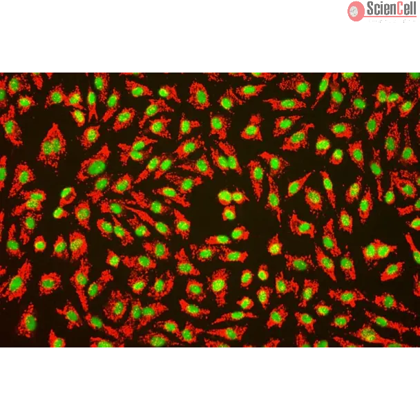
In senescent endothelial cells, DS2-sourced indole-3-lactic acid (ILA) activated aryl hydrocarbon receptor (AhR) signaling. This activation reduced the expression of senescence markers (p16, γ-H2A.X) and decreased the levels of pro-inflammatory molecules (ICAM-1, VCAM-1). In aged mice, DS2 supplementation increased the abundance of beneficial bacteria, including Lactobacillus and Ligilactobacillus. DS2 administration also elevated plasma ILA and IL-22 levels, and reduced intestinal permeability. This was evidenced by the expansion of IL-22-producing type 3 innate lymphoid cells (ILC3s) and activation of the AhR-IL-22 axis. Consequently, DS2 enhanced intestinal barrier integrity and mitigated systemic inflammation (TNF-α, IL-6). Exogenous ILA was sufficient to recapitulate these benefits by potentiating gut mucosal immunity and attenuating inflammaging, as all these effects were abolished by the AhR antagonist CH223191.
Conclusions
We demonstrate that DS2 mitigates inflammaging by producing ILA, which acts as a key metabolite to activate the AhR-IL-22-ILC3 axis. Our findings highlight the potential of targeting the gut-immune axis with specific probiotics as a novel strategy against age-related immune decline. Less
Chronic kidney disease (CKD) is associated with inflammation and cardiovascular complications and is partly exacerbated by the uremic toxin indoxyl sulfate (IS). IS is kn... More
Chronic kidney disease (CKD) is associated with inflammation and cardiovascular complications and is partly exacerbated by the uremic toxin indoxyl sulfate (IS). IS is known to activate the aryl hydrocarbon receptor (AhR) to promote vascular inflammation. On the other hand, avenanthramide C (Ave), an oat-derived polyphenol, has antioxidative and anti-inflammatory properties. Therefore, we investigated whether Ave can suppress IS-induced inflammatory responses. Less
In previous work we discovered that T lymphocytes play a prominent role in the rise of brain metastases of ER-negative breast cancers. In the present study we explored ho... More
In previous work we discovered that T lymphocytes play a prominent role in the rise of brain metastases of ER-negative breast cancers. In the present study we explored how T lymphocytes promote breast cancer cell penetration through the blood brain barrier (BBB). An in vitro BBB model was employed to study the effects of T lymphocytes on BBB trespassing capacity of three different breast carcinoma cell lines. Differential protein expression was explored by comparing the proteomes of the breast cancer cells before and after co-culture with activated T lymphocytes using liquid chromatography-mass spectrometry (LC-MS). siRNA was used to silence protein expression in the breast cancer cells to study contribution to in vitro BBB passage. Furthermore, protein expression in primary breast cancer tissues was explored and related to brain-metastatic potential. Co-culturing with activated T lymphocytes or their conditioned medium (CM) resulted in increased passage through the in vitro BBB. The effects were less for cell line MDA-MB-231-B2M2 (brain affinity) as compared to MDA-MB-231 and SK-BR-7. Mass spectrometry-based proteomics revealed significant alterations in the expression of 35 proteins by the breast cancer cell lines upon T cell contact. Among the proteins is coronin-1 A, a protein related to cell motility. Knockdown of CORO1A in the breast cancer cells reduced their ability to cross the artificial BBB to 60%. The effects were significantly less for the cell line derived from breast cancer with affinity for brain. The expression of coronin-1A was confirmed by immunohistochemistry and RT-PCR of 52 breast cancer samples of patients with metastasized breast cancers, with and without brain locations. Lastly, CORO1A upregulation was validated in a publicly available mRNA expression database from 204 primary breast cancers with known metastatic sites. We conclude that T lymphocytes trigger cancer cells to express proteins including coronin-1A that enable the cancer cells to cross an in vitro BBB. In addition, a prominent role of coronin-1A in the formation of cerebral metastases in breast cancer patients is strongly suggestive by its upregulation in tissue samples of breast cancer patients with brain metastases. Less
Background Achieving a stable vasculature is crucial for tissue regeneration. Endothelial cells initiate vascular morphogenesis, followed by mural cells that stabilize ne... More
Background Achieving a stable vasculature is crucial for tissue regeneration. Endothelial cells initiate vascular morphogenesis, followed by mural cells that stabilize new vessels. This study investigates the in vivo effects of Sema4D-Plexin-B1 signaling on stem cells from human exfoliated deciduous teeth (SHED)-supported angiogenesis, focusing on its mechanism in PDGF-BB secretion. We also explore macrophages as an endogenous source of Sema4D for vascular stabilization. Methods The in vivo Matrigel plug angiogenesis assay was conducted to examine the impact of Sema4D on vessel formation and stabilization supported by SHED. Knockdown of Plexin-B1 in human umbilical vein endothelial cells (HUVECs) and the application of PDGFR-β inhibitors were utilized to explore the fundamental regulatory mechanisms. Furthermore, the m6A methylation levels of total RNA and the expression of Methyltransferase-like 3 (METTL3) were assessed under conditions of Sema4D treatment in vitro. An ELISA was employed to measure the levels of Sema4D in the supernatants derived from THP-1 cell-mediated macrophages. Additionally, a three-dimensional vasculature-on-a-chip microfluidic device was employed to investigate the role of M2c macrophage-derived Sema4D in the stabilization of vascular structures. Results Sema4D induced the formation of a greater number of perfused vessels by HUVECs and enhanced the coverage of these vessels by SM22α-positive SHED (SM22α+SHED). Conversely, the knockdown of the Plexin-B1 receptor in HUVECs or inhibition of PDGFR-β reversed the Sema4D-induced vascular stabilization, thereby confirming the regulatory role of the Plexin-B1/PDGF-BB axis in the recruitment of mural cells mediated by Sema4D. Mechanistically, Sema4D was found to upregulate the expression of methyltransferases, specifically METTL3, and to elevate the level of m6A modification in HUVECs. This modification was determined to be critical for enhancing PDGF-BB secretion, suggesting that Sema4D activates an epigenetic regulatory mechanism. Additionally, we investigated the secretion of Sema4D by various macrophage phenotypes, identifying that M2c macrophages secrete significant levels of Sema4D. This secretion recruits SM22α+SHED as mural cells by inducing endothelial PDGF production on a vasculature-on-a-chip platform, indicating a potential role for macrophages in facilitating vascular stabilization. Conclusions Sema4D acts on Plexin-B1, inducing METTL3-mediated PDGF-BB secretion to recruit SHED to stabilize vessels. Macrophages could be a key source of Sema4D for vascular stabilization. Less
Angiogenesis is a significant pathogenic characteristic of diabetic microangiopathy. Advanced glycation end products (AGEs) are considerably elevated in diabetic tissues ... More
Angiogenesis is a significant pathogenic characteristic of diabetic microangiopathy. Advanced glycation end products (AGEs) are considerably elevated in diabetic tissues and can affect vascular endothelial cell shape and function. Regulation of the vascular endothelial growth factor (VEGF)-VEGF receptor 2 (VEGFR2) signaling pathway is a critical mechanism in the regulation of angiogenesis, and VEGFR2 activity can be modified by post-translational changes. However, little research has been conducted on the control of small ubiquitin-related modifier (SUMO)-mediated VEGFR2 alterations. The current study investigated this using human umbilical vein endothelial cells (HUVECs) in conjunction with immunoblotting and immunofluorescence. AGEs increased Nrf2 translocation to the nucleus and promoted VEGFR2 expression. They also increased the expression of sentrin/SUMO-specific protease 6 (SENP6), which de-SUMOylated VEGFR2, and immunofluorescence indicated a reduction in VEGFR2 accumulation in the Golgi and increased VEGFR2 transport from the Golgi to the cell membrane surface via the coatomer protein complex subunit beta 2. VEGFR2 on the cell membrane was linked to VEGF generated by pericytes, triggering the VEGF signaling cascade. In conclusion, this study demonstrates that SENP6 regulates VEGFR2 trafficking from the Golgi to the endothelial cell surface. The SENP6-VEGFR2 pathway plays a critical role in pathological angiogenesis. Less
Trans, trans-2,4-decadienal (tt-DDE) is a highly reactive unsaturated aldehyde that widely present in foods. This study aimed to investigate the toxic effects of tt-DDE e... More
Trans, trans-2,4-decadienal (tt-DDE) is a highly reactive unsaturated aldehyde that widely present in foods. This study aimed to investigate the toxic effects of tt-DDE exposure on human umbilical vein endothelial cells (HUVECs) and the possible protective effects of green tea catechins. tt-DDE exposure induced pyroptosis and NLRP3 inflammasome activation in endothelial cells, as evidenced by increased LDH release and PI-positive cells, and elevated protein expressions of NLRP3, cleaved caspase-1, and GSDMD-N. NLRP3 inhibitor (MCC950) efficiently suppressed the tt-DDE-induced NLRP3 inflammasome activation and pyroptosis. Additionally, epigallocatechin-3-gallate (EGCG) showed the strongest activity among the four green tea catechins, which significantly alleviated tt-DDE-induced cytotoxicity. Moreover, EGCG effectively attenuated tt-DDE-induced endothelial cell pyroptosis and dysfunction by inhibiting NLRP3 inflammasome. Results of cultured mesenteric arteries further confirmed that EGCG prevented tt-DDE-induced endothelial dysfunction and pyroptosis. These results provide novel insights into tt-DDE-induced endothelial injury, and demonstrate the protective role of EGCG against tt-DDE-associated endothelial toxicity. Less
Biodegradable shape memory polymers (SMP) with suitable transition temperatures (Tr) and mechanical properties are highly demanded in biomedical field as deployable medic... More
Biodegradable shape memory polymers (SMP) with suitable transition temperatures (Tr) and mechanical properties are highly demanded in biomedical field as deployable medical devices. Herein, we report a 4D printing shape memory Inferior Vena Cava Filters (IVCFs), an implantation device, which could prevent the fatal pulmonary embolism, to exemplify the applicability of the biodegradable shape memory polymer in biomedical device field. The IVCF composed of poly(glycerol sebacate) acrylate-co-hydroxyethyl methacrylate (PGSA-co-HEMA) was digital light processing (DLP) 3D printed. The appropriate mechanical property and Tr = 37.8 °C, which is close to human body temperature, was tailored by tuning the ratio of the raw material. PGSA-PHEMA presents an excellent cytocompatibility, hemocompatibility and histocompatibility as implants. Besides, in vitro degradation results indicate the biodegradability but withhold the mechanical properties within the service time. Furthermore, the simulated filter deploying and fully emboli interception verifies the successful realization of the concept of rapid, minimally invasive and controllable implantation of the 4D printing of IVCFs through the SMP transformation process, and the feasibility of the filter as well. Therefore, this work provides a new biocompatible SMP and offers a new strategy for developing deployable medical devices. Less
Group 1 innate lymphoid cells (ILCs) comprise a heterogeneous family of cytotoxic natural killer (NK) cells and ILC1s. We identify a population of “liver-type” ILC1s ... More
Group 1 innate lymphoid cells (ILCs) comprise a heterogeneous family of cytotoxic natural killer (NK) cells and ILC1s. We identify a population of “liver-type” ILC1s with transcriptional, phenotypic, and functional features distinct from those of conventional and liver-resident NK cells as well as from other previously described human ILC1 subsets. LT-ILC1s are CD49a+CD94+CD200R1+, express the transcription factor T-BET, and do not express the activating receptor NKp80 or the transcription factor EOMES. Similar to NK cells, liver-type ILC1s produce IFN-γ, TNF-α, and GM-CSF; however, liver-type ILC1s also produce IL-2 and lack perforin and granzyme-B. Liver-type ILC1s are expanded in cirrhotic liver tissues, and they can be produced from blood-derived ILC precursors in vitro in the presence of TGF-β1 and liver sinusoidal endothelial cells. Cells with similar signature and function can also be found in tonsil and intestinal tissues. Collectively, our study identifies and classifies a population of human cross-tissue ILC1s. Less
Severe bone trauma can lead to poor or delayed bone healing and nonunion. Bone regeneration is based on the interaction between osteogenesis and angiogenesis. Angiogenesi... More
Severe bone trauma can lead to poor or delayed bone healing and nonunion. Bone regeneration is based on the interaction between osteogenesis and angiogenesis. Angiogenesis serves a unique role in the repair and remodeling of bone defects. Monocyte chemoattractant protein‑1, also known as CC motif ligand 2 (CCL2), is a member of the CC motif chemokine family and was the first human chemokine to be revealed to be an effective chemokine of monocytes. However, its underlying mechanism in angiogenesis of bone defect repair remains to be elucidated. Therefore, the present study investigated the detailed mechanism by which CCL2 promoted angiogenesis in bone defects based on cell and animal model experiments. In the present study, CCL2 promoted proliferation, migration and tube formation in human umbilical vein endothelial cells (HUVECs) in a concentration‑dependent manner. Western blot analysis revealed that treatment of HUVECs with CCL2 upregulated the protein expression levels of rho‑associated coiled‑coil‑containing protein kinase (Rock)1, Rock2, N‑cadherin, c‑Myc and VEGFR2. Furthermore, CCL2 promoted the expression of MAPK/ERK1/2/MMP9, PI3K/AKT and Wnt/β‑catenin signaling pathway‑related proteins, which also demonstrated that CCL2 promoted these functions in HUVECs. Immunohistochemical staining of Sprague Dawley rat femurs following bone defects revealed that VEGF expression was positive in the newly formed bone area in each group, while the expression area of VEGF in the CCL2 addition group was markedly increased. Therefore, CCL2 is a potential therapeutic approach for bone defect repair and reconstruction through the mechanism of angiogenesis‑osteogenesis coupling. Less
Background
Distraction osteogenesis (DO) is an efficacious but lengthy procedure to reconstruct segmental bone defects under the principle of tension-stress, during which... More
Background
Distraction osteogenesis (DO) is an efficacious but lengthy procedure to reconstruct segmental bone defects under the principle of tension-stress, during which the periosteum-mediated mechanical stimulation plays a pivotal role. Inspired by the dynamic process of DO and the mechanical stimulation of periosteum, a new design of bionic periosteum was developed to simulate the mechanical transduction of natural periosteum for the application in DO procedure.
Methods
In this study, an injectable organic-inorganic hybrid hydrogel was developed based on a novel combination of the PEGylated poly (glycerol sebacate) (PEGS) polymer network and in situ formed CaP nanoparticles (ICPNs). Rat bone marrow mesenchymal stem cells (rBMSCs) and human umbilical vein endothelial cells (HUVECs) were cultured and tested in vitro to evaluate biocompatibility, cell adhesion, proliferation, and pro-osteogenic and pro-angiogenic activity. In vivo experiments were conducted in the rat tibial model of distraction osteogenesis.
Results
The developed nanocomposite hydrogels exhibited excellent injectability, robust bone adhesion, superior stretchability, and enhanced osteogenic activity. The results of in vitro and in vivo studies showed that PEGS/ICPN hydrogels could promote new bone formation and mineralization during the dynamic distraction process through the synergistic effects of angiogenesis and osteogenesis.
Conclusions
This periosteum-inspired nanocomposite hydrogel represents a mechanobiology approach for effectively restoring large bone defects through the dynamic DO process. Less
Inhibition of angiogenesis is considered as one of the desirable pathways for the treatment of tumor growth and metastasis. Herein we demonstrated that a series of pyridi... More
Inhibition of angiogenesis is considered as one of the desirable pathways for the treatment of tumor growth and metastasis. Herein we demonstrated that a series of pyridinyl-thiazolyl carboxamide derivatives were designed, synthesized and examined against angiogenesis through a colony formation and migration assays of human umbilical vein endothelial cells (HUVECs) in vitro. A structure-activity relationship (SAR) study was carried out and optimization toward this series of compounds resulted in the discovery of N-(3-methoxyphenyl)-4-methyl-2-(2-propyl-4-pyridinyl)thiazole-5-carboxamide (3k). The results indicated that compound 3k showed similar or better effects compared to Vandetanib in suppressing HUVECs colony formation and migration as well as VEGF-induced angiogenesis in the aortic ring spreading model and chick embryo chorioallantoic membrane (CAM) model. More importantly, compound 3k also strongly blocked tumor growth with the dosage of 30 mg/kg/day, and subsequent mechanism exploration suggested that this series of compounds took effect mainly through angiogenesis signaling pathways. Together, these results suggested compound 3k may serve as a lead for a novel class of angiogenesis inhibitors for cancer treatments. Less
Ectopic apoptosis of vascular cells plays a critical role in the early stage development of diabetic retinopathy (DR). Uncoupling protein 2 (UCP2) is a mitochondrial modu... More
Ectopic apoptosis of vascular cells plays a critical role in the early stage development of diabetic retinopathy (DR). Uncoupling protein 2 (UCP2) is a mitochondrial modulator which protects against endothelial dysfunction. However, the role which UCP2 plays in endothelial apoptosis and its association with DR was unclear. In the present study, we investigated whether UCP2 functioned as an inhibitor of DR in endothelial cells. Firstly, we noted that in UCP2‑knockout mice retinal cell death and damage in vivo was similar to that of db/db diabetic mice. Additionally, UCP2 knockdown induced caspase-3 activation and exaggerated high glucose (HG)-induced apoptosis of human umbilical vein endothelial cells (HUVECs). Conversely, adenovirus-mediated UCP2 overexpression inhibited the apoptosis of HUVECs and HG-induced caspase-3 activation. Furthermore, HG treatment resulted in the opening of the permeability transition pore (PTP) and liberation of cytochrome c from mitochondria to the cytosol in HUVECs. Notably, UCP2 overexpression inhibited these processes. Furthermore, adenovirus-mediated UCP2 overexpression led to a significant increase in intracellular nitric oxide (NO) levels and a decrease in reactive oxygen species (ROS) generation in HUVECs. Collectively, these data suggest that UCP2 plays an anti-apoptotic role in endothelial cells. Thus, we suggest that approaches which augment UCP2 expression in vascular endothelial cells aid in preventing the early stage development and progression of DR. Less
Background Endothelial cells (ECs) form blood vessels through angiogenesis that is regulated by coordination of vascular endothelial growth factor (VEGF), Notch, transfor... More
Background Endothelial cells (ECs) form blood vessels through angiogenesis that is regulated by coordination of vascular endothelial growth factor (VEGF), Notch, transforming growth factor β, and other signals, but the detailed molecular mechanisms remain unclear. Methods and Results Small RNA sequencing initially identified miR‐342‐5p as a novel downstream molecule of Notch signaling in ECs. Reporter assay, quantitative reverse transcription polymerase chain reaction and Western blot analysis indicated that miR‐342‐5p targeted endoglin and modulated transforming growth factor β signaling by repressing SMAD1/5 phosphorylation in ECs. Transfection of miR‐342‐5p inhibited EC proliferation and lumen formation and reduced angiogenesis in vitro and in vivo, as assayed by using a fibrin beads–based sprouting assay, mouse aortic ring culture, and intravitreal injection of miR‐342‐5p agomir in P3 pups. Moreover, miR‐342‐5p promoted the migration of ECs, accompanied by reduced endothelial markers and increased mesenchymal markers, indicative of increased endothelial–mesenchymal transition. Transfection of endoglin at least partially reversed endothelial–mesenchymal transition induced by miR‐342‐5p. The expression of miR‐342‐5p was upregulated by transforming growth factor β, and inhibition of miR‐342‐5p attenuated the inhibitory effects of transforming growth factor β on lumen formation and sprouting by ECs. In addition, VEGF repressed miR‐342‐5p expression, and transfection of miR‐342‐5p repressed VEGFR2 and VEGFR3 expression and VEGF‐triggered Akt phosphorylation in ECs. miR‐342‐5p repressed angiogenesis in a laser‐induced choroidal neovascularization model in mice, highlighting its clinical potential. Conclusions miR‐342‐5p acts as a multifunctional angiogenic repressor mediating the effects and interaction among angiogenic pathways. Less
Previous studies have demonstrated that Smyd1 plays a critical role in cardiomyocyte differentiation, cardiac morphogenesis and myofibril organization. In this study, we ... More
Previous studies have demonstrated that Smyd1 plays a critical role in cardiomyocyte differentiation, cardiac morphogenesis and myofibril organization. In this study, we uncovered a novel function of Smyd1 in the regulation of endothelial cells (ECs). Our data showed that Smyd1 is expressed in vascular endothelial cells, and knockdown of SMYD1 in endothelial cells impairs EC migration and tube formation. Furthermore, Co-IP and GST pull-down assays demonstrated that SMYD1 is associated with the Serum Response Factor (SRF). EMSA assays further showed that SMYD1 forms a complex with SRF and enhances SRF DNA binding activity. Our studies indicate that SMYD1 serves as an SRF-interacting protein, enhances SRF DNA binding activity, and is required for EC migration and tube formation to regulate angiogenesis. Less
Spinal cord injury (SCI) induces the disruption of the blood-spinal cord barrier (BSCB) which leads to infiltration of blood cells, an inflammatory response, and neuronal... More
Spinal cord injury (SCI) induces the disruption of the blood-spinal cord barrier (BSCB) which leads to infiltration of blood cells, an inflammatory response, and neuronal cell death, resulting spinal cord secondary damage. Retinoic acid (RA) has a neuroprotective effect in both ischemic brain injury and SCI, however the relationship between BSCB disruption and RA in SCI is still unclear. In this study, we demonstrated that autophagy and ER stress are involved in the protective effect of RA on the BSCB. RA attenuated BSCB permeability and decreased the loss of tight junction (TJ) molecules such as P120, β-catenin, Occludin and Claudin5 after injury in vivo as well as in Brain Microvascular Endothelial Cells (BMECs). Moreover, RA administration improved functional recovery in the rat model of SCI. RA inhibited the expression of CHOP and caspase-12 by induction of autophagic flux. However, RA had no significant effect on protein expression of GRP78 and PDI. Furthermore, combining RA with the autophagy inhibitor chloroquine (CQ) partially abolished its protective effect on the BSCB via exacerbated ER stress and subsequent loss of tight junctions. Taken together, the neuroprotective role of RA in recovery from SCI is related to prevention of of BSCB disruption via the activation of autophagic flux and the inhibition of ER stress-induced cell apoptosis. These findings lay the groundwork for future translational studies of RA for CNS diseases, especially those related to BSCB disruption. Less
Numerous epidemiological studies have shown that subclinical hypothyroidism (SCH) can impair endothelial function and cause dyslipidemia. Studies have evaluated the effec... More
Numerous epidemiological studies have shown that subclinical hypothyroidism (SCH) can impair endothelial function and cause dyslipidemia. Studies have evaluated the effects of thyroid stimulating hormone (TSH) on endothelial cells, but the mechanism underlying the proatherosclerotic effect of increased TSH levels remains unclear. In the present study, SCH rat models were established in thyroidectomized Wistar rats that were given l-T4 daily. The results showed that in vivo, the expression of osteopontin (OPN) vascular cell adhesion molecule (VCAM-1), and levels of integrin αvβ3 in the aortic tissue in SCH and Hypothyroidism (CH) groups was higher than in the control group. However, the effect in the SCH group was higher than in the CH group. In vitro, results showed that different concentration and time gradients of TSH stimulation could increase the expression of OPN, VCAM-1, and integrin αvβ3, and this was accompanied by extracellular signal regulated kinase 1/2 (Erk1/2) and Akt activation in human umbilical vein endothelial cells (HUVECs). TSH induced elevation of these proatherosclerotic factors was partially suppressed by a specific Akt inhibitor but not by a specific Erk inhibitor. Findings suggested that the endothelial dysfunction caused by SCH was related to increased proatherosclerotic factors induced by TSH via Akt activation. Less
Background: Mesenchymal stem cells derived from human umbilical cord tissue, termed UCX®, have the potential to promote a full range of events leading to tissue regenera... More
Background: Mesenchymal stem cells derived from human umbilical cord tissue, termed UCX®, have the potential to promote a full range of events leading to tissue regeneration and homeostasis. The main goal of this work was to investigate UCX® action in experimentally induced hindlimb ischemia (HLI).
Methods: UCX®, obtained by using a proprietary technology developed by ECBio (Amadora, Portugal), were delivered via intramuscular injection to C57BL/6 females after unilateral HLI induction. Perfusion recovery, capillary and collateral density increase were evaluated by laser doppler, CD31 immunohistochemistry and diaphonisation, respectively. The activation state of endothelial cells (ECs) was analysed after EC isolation by laser capture microdissection microscopy followed by RNA extraction, cDNA synthesis and quantitative RT-PCR analysis. The UCX®-conditioned medium was analysed on Gallios flow cytometer. The capacity of UCX® in promoting tubulogenesis and EC migration was assessed by matrigel tubule formation and wound-healing assay, respectively.
Results: We demonstrated that UCX® enhance angiogenesis in vitro via a paracrine effect. Importantly, after HLI induction, UCX® improve blood perfusion by stimulating angiogenesis and arteriogenesis. This is achieved through a new mechanism in which durable and simultaneous upregulation of transforming growth factor β2, angiopoietin 2, fibroblast growth factor 2, and hepatocyte growth factor, in endothelial cells is induced by UCX®.
Conclusions: In conclusion, our data demonstrate that UCX® improve the angiogenic potency of endothelial cells in the murine ischemic limb suggesting the potential of UCX® as a new therapeutic tool for critical limb ischemia.
Keywords: Angiogenesis; Arteriogenesis; Critical limb ischemia; Endothelial cells; Hindlimb ischemia; Mesenchymal stem cells; UCX®. Less
It has been shown that forced expression of four mouse stem cell factors (OCT4, Sox2, Klf4, and c-Myc) changed the phenotype of rat endothelial cells to vascular progenit... More
It has been shown that forced expression of four mouse stem cell factors (OCT4, Sox2, Klf4, and c-Myc) changed the phenotype of rat endothelial cells to vascular progenitor cells. The present study aimed to explore whether the expression of OCT4 alone might change the phenotype of human umbilical vein endothelial cells (HUVECs) to endothelial progenitor cells and, if so, to examine the possible mechanism involved. A Matrigel-based in vitro angiogenesis assay was used to
evaluate the angiogenesis of the cells; the gene expression profile was analyzed by an oligonucleotide probe-based gene array chip and validated by RT-QPCR. The cellular functions of the mRNAs altered by OCT4 were analyzed with Gene Ontology. We found that induced ectopic expression of mouse OCT4 in HUVECs significantly enhanced angiogenesis of the cells, broadly changed the gene expression profile and particularly increased the expression of CD133, CD34, and VEGFR2 (KDR) which are characteristic marker molecules for endothelial progenitor cells (EPCs). Furthermore by analyzing the cellular functions that were targeted by the mRNAs altered by OCT4 we found that stem cell maintenance and cell differentiation were among the top functional response targeted by up-regulated and down-regulated mRNAs upon forced expression of OCT4. These results support the argument that OCT4 remodels the phenotype of HUVECs from endothelial cells to EPCs by up-regulating the genes responsible for stem cell maintenance and down-regulating the genes for cell differentiation.
Key words: Endothelial Progenitor Cells; Human Umbilical Vein Endothelial Cells; Angiogenesis; Gene Expression; Octamer-binding transcription factor 4. Less
Background: Dysfunction of vascular endothelium is implicated in many pathological situations. Cytoskeleton plays an importance role in vascular endothelial permeability ... More
Background: Dysfunction of vascular endothelium is implicated in many pathological situations. Cytoskeleton plays an importance role in vascular endothelial permeability barrier and inflammatory response. Many Chinese herbs have the endothelial protective effect, of which, "Astragalus membranaceus" is a highly valued herb for treatment of cardiovascular and renal diseases in traditional Chinese medicine, In this study, we tested whether calycosin-7-O-β-D-glucoside (Calycosin), a main effective monomer component of "Astragalus membranaceus", could protect endothelial cells from bacterial endotoxin (LPS)-induced cell injury. Less
EXPERIMENTAL AND THERAPEUTIC MEDICINE 10: 1404-1412, 20151404 Abstract. The aim of the present study was to examine the mechanisms through which fenofibrate inhibits the ... More
EXPERIMENTAL AND THERAPEUTIC MEDICINE 10: 1404-1412, 20151404 Abstract. The aim of the present study was to examine the mechanisms through which fenofibrate inhibits the ability of human retinal pigment epithelial cells (RPE cells) exposed to hypoxia to stimulate the proliferation and migration of human umbilical vein endothelial cells (HUVECs). For this purpose, RPE cells and HUVECs were divided into the following groups: RPE-normoxia, RPE + fenofibrate, RPE-hypoxia, RPE hypoxia + fenofibrate; HUVECs normal culture and HUVECs + RPE-hypoxia culture supernatant. RPE cell hypoxia was induced by cobalt(II) chloride (CoCl2). A superoxide anion probe was used to measure the production of superoxide anion, which is indicative of hypoxic conditions. Cell proliferation was assessed by MTT assay, and the expression of vascular endothelial growth factor C (VEGFC) and vascular endothelial growth factor receptor-3 (VEGFR-3) in the RPE cell culture supernatant was measured by enzyme-linked immunosorbent assay (ELISA). The migration ability of the HUVECs was determined by scratch-wound assay, and the angiogenic ability of the HUVECs was examined by measuring cell lumen forma- tion. The mRNA and protein expression levels of VEGFC and VEGFR-3 in the RPE cells were measured by RT-qPCR and western blot analysis, respectively. Our results revealed that fenofibrate inhibited the increase in the expression and release of VEGFC and VEGFR-3 into the RPE cell culture superna- tant induced by exposure to hypoxia. The culture of HUVECs in medium supernatant of RPE cells epxosed to hypoxia enhanced the viability and migration ability of the HUVECs and promoted lumen formation; these effects were inhibited by fenofibrate. In conclusion, our data demonstrated that the exposure of RPE cells to hypoxia induced the expression and release of VEGFC and VEGFR-3 into the cell culture super- natant. The culture of HUVECs in conditioned medium from RPE cells exposed to hypoxia increased VEGFC and VEGFR-3 expression, and promoted the proliferation and migration of the HUVECs, as well as capillary tube formation, suggesting that RPE cells play an important role in the formation of choroidal neovascularization resulting from hypoxia. Fenofibrate inhib- ited the upregulation of VEGFC and VEGFR-3 in the RPE cells exposed to hypoxia, and thus reduced the ability of HUVECs to form new blood vessels. Less
Endothelial senescence plays crucial roles in diabetic vascular complication. Recent evidence indicated that transient hyperglycaemia could potentiate persistent diabetic... More
Endothelial senescence plays crucial roles in diabetic vascular complication. Recent evidence indicated that transient hyperglycaemia could potentiate persistent diabetic vascular complications, a phenomenon known as “metabolic memory.” Although SIRT1 has been demonstrated to mediate high glucose-induced endothelial senescence, whether and how “metabolic memory” would affect endothelial senescence through SIRT1 signaling remains largely unknown. In this study, we investigated the involvement of SIRT1 axis as well as the protective effects of resveratrol (RSV) and metformin (MET), two potent SIRT1 activators, during the occurrence of “metabolic memory” of cellular senescence (senescent “memory”). Human umbilical vascular endothelial cells (HUVECs) were cultured in either normal glucose (NG)/high glucose (HG) media for 6 days, or 3 days of HG followed by 3 days of NG (HN), with or without RSV or MET treatment. It was shown that HN incubation triggered persistent downregulation of deacetylase SIRT1 and upregulation of acetyltransferase p300, leading to sustained hyperacetylation (at K382) and activation of p53, and subsequent p53/p21-mediated senescent “memory.” In contrast, senescent “memory” was abrogated by overexpression of SIRT1 or knockdown of p300. Interestingly, we found that SIRT1 and p300 could regulate each other in response to HN stimulation, suggesting that a delicate balance between acetyltransferases and deacetylases may be particularly important for sustained acetylation and activation of non-histone proteins (such as p53), and eventually the occurrence of “metabolic memory.” Furthermore, we found that RSV or MET treatment prevented senescent “memory” by modulating SIRT1/p300/p53/p21 pathway. Notably, early and continuous treatment of MET, but not RSV, was particularly important for preventing senescent “memory.” In conclusion, short-term high glucose stimulation could induce sustained endothelial senescence via SIRT1/p300/p53/p21 pathway. RVS or MET treatment could enhance SIRT1-mediated signaling and thus protect against senescent “memory” independent of their glucose lowering mechanisms. Therefore, they may serve as promising therapeutic drugs against the development of “metabolic memory.” Less
The success of bioengineered dental pulp depends on two principles, (1) whether the transplanted tissue can develop its own vascular endothelial tubule network and (2) wh... More
The success of bioengineered dental pulp depends on two principles, (1) whether the transplanted tissue can develop its own vascular endothelial tubule network and (2) whether the host vasculature can be induced to penetrate the bioengineered pulp replacement and conjoin. Major inductive molecules that participate in laying down blood vessels include vascular endothelial growth factor (VEGF), ephrinB2, and hypoxia-inducible factor 1α (HIF-1α). Being able to modulate the genes encoding these angiogenic molecules is a therapeutic target in pulp regeneration for endogenous blood vessel formation, prevention of graft rejection, and exclusion of infection. Once implanted inside the root canal, bioengineered pulp is subjected to severe hypoxia that causes tissue degeneration. However, short-term hypoxia is known to stimulate angiogenesis. Thus, it may be feasible to prime dental cells for angiogenic activity before implantation. Stem cells from apical papilla (SCAP) are arguably one of the most potent and versatile dental stem cell populations for bioengineering pulp in vitro. Our study aimed to investigate whether coculture of SCAP and human umbilical vein endothelial cells (HUVECs) under hypoxia promotes the formation of endothelial tubules and a blood vessel network. In addition, we clarified the interplay between the genes that orchestrate these important angiogenic molecules in SCAP under hypoxic conditions. We found that SCAP cocultured with HUVEC at a 1:5 ratio increased the number of endothelial tubules, tubule lengths, and branching points. Fluorescence staining showed that HUVEC formed the trunk of tubular structures, whereas SCAP located adjacent to the endothelial cell line, resembling the pericyte location. When we used CoCl2 (0.5 mM) to induce hypoxic environment, the expression of proteins, HIF-1α and VEGF, and transcript of ephrinB2 in SCAP was upregulated. However, minimal VEGF levels in supernatants of HUVEC and coculture Petri dishes were detected, suggesting that VEGF secreted by SCAP might be used by HUVEC to accelerate the formation of vessel-like structures. Taken together, we revealed that artificial hypoxia stimulates angiogenic responses in SCAP for possible use in engineering dental pulp replacements. Our results may help to delineate the optimal therapeutic target to promote angiogenesis so that future bioengineered pulp replacements integrate faster and permanently within the host. Less
To investigate the effect of endostar on specific angiogenesis induced by human hepatocellular carcinoma, this research systematically elucidated the inhibitory effect on... More
To investigate the effect of endostar on specific angiogenesis induced by human hepatocellular carcinoma, this research systematically elucidated the inhibitory effect on HepG2-induced angiogenesis by endostar from 50 ng/mL to 50000 ng/mL. We employed fluorescence quantitative Boyden chamber analysis, wound-healing assay, flow cytometry examination using a coculture system, quantitative analysis of tube formation, and in vivo Matrigel plug assay induced by HCC conditioned media (HCM) and HepG2 compared with normal hepatocyte conditioned media (NCM) and L02. Then, we found that endostar as a tumor angiogenesis inhibitor could potently inhibit human umbilical vein endothelial cell (HUVEC) migration in response to HCM after four- to six-hour action, inhibit HCM-induced HUVEC migration to the lesion part in a dose-dependent manner between 50 ng/mL and 5000 ng/mL at 24 hours, and reduce HUVEC proliferation in a dose-dependent fashion. Endostar inhibited HepG2-induced tube formation of HUVECs which peaked at 50 ng/mL. In vivo Matrigel plug formation was also significantly reduced by endostar in HepG2 inducing system rather than in L02 inducing system. It could be concluded that, at cell level, endostar inhibited the angiogenesis-related biological behaviors of HUVEC in response to HCC, including migration, adhesion proliferation, and tube formation. At animal level, endostar inhibited the angiogenesis in response to HCC in Matrigel matrix. Less
Cigarette smoking, a major independent risk factor of atherosclerosis, can cause oxidative and inflammatory damage of vascular tissue. Heme oxygenase-1 (HO-1) is an endog... More
Cigarette smoking, a major independent risk factor of atherosclerosis, can cause oxidative and inflammatory damage of vascular tissue. Heme oxygenase-1 (HO-1) is an endogenous cytoprotective enzyme with an anti-oxidant role in cells. The aim of the present study was to investigate whether HO-1 was able to protect vascular and endothelial cells from the oxidative damage induced by cigarette smoking. It was observed that cigarette smoking was able to induce the generation of the reactive oxygen species (ROS) in carotid arteries of rats. Hemin, a widely used HO-1 inducer, was able to reduce the generation of ROS. In addition, when human umbilical vein endothelial cells (HUVECs) were cultured in the serum of smoking rats, this was able to increase ROS, and the protective effect of hemin was also observed in this system. In conclusion, the present study demonstrated that cigarette smoking causes oxidative damage of vascular cells and HUVECs by inducing the generation of ROS, while HO-1 has an anti-oxidant effect in this course. This also implied that hemin, an inducer of HO-1, may have potential therapeutic applicability in the prevention of vascular diseases caused by cigarette smoking. Less
Macrophage invasion is an important event during arteriogenesis, but the underlying mechanism is still only partially understood. The present study tested the hypothesis ... More
Macrophage invasion is an important event during arteriogenesis, but the underlying mechanism is still only partially understood. The present study tested the hypothesis that nitric oxide (NO) and VE-cadherin, two key mediators for vascular permeability, contribute to this event in a rat ischemic hindlimb model. In addition, the effect of NO on expression of VE-caherin and endothelial permeability was also studied in cultured HUVECs. We found that: 1) in normal arteriolar vessels (NAV), eNOS was moderately expressed in endothelial cells (EC) and iNOS was rarely detected. In contrast, in collateral vessels (CVs) induced by simple femoral artery ligation, both eNOS and iNOS were significantly upregulated (P<0.05). Induced iNOS was found mainly in smooth muscle cells, but also in other vascular cells and macrophages; 2) in NAV VE-cadherin was strongly expressed in EC. In CVs, VE-cadherin was significantly downregulated, with a discontinuous and punctate pattern. Administration of nitric oxide donor DETA NONOate (NONOate) further reduced the amounts of Ve-cadherin in CVs, whereas NO synthase inhibitor L-NAME inhibited downregulation of VE-cadherin in CVs; 3) in normal rats Evans blue extravasation (EBE) was low in the musculus gracilis, FITC-dextron leakage was not detected in the vascular wall and few macrophages were observed in perivascular space. In contrast, EBE was significantly increased in femoral artery ligation rats, FITC-dextron leakage and increased amounts of macrophages were detected in CVs, which were further enhanced by administration of NONOate, but inhibited by L-NAME supplement; 4) in vitro experiments confirmed that an increase in NO production reduced VE-cadherin expression, correlated with increases in the permeability of HUVECs. In conclusion, our data for the first time reveal the expression profile of VE-cadherin and alterations of vascular permeability in CVs, suggesting that NO-mediated VE-cadherin pathway may be one important mechanism responsible, at least in part, for macrophage invasion during arteriogenesis. Less
Angiogenesis plays a critical role in the growth and metastasis of tumors, which makes it an attractive target for anti-tumor drug development. Deoxypodophyllotoxin (DPT)... More
Angiogenesis plays a critical role in the growth and metastasis of tumors, which makes it an attractive target for anti-tumor drug development. Deoxypodophyllotoxin (DPT), a natural product isolated from Anthriscus sylvestris, inhibits cell proliferation and migration in various cancer cell types. Our previous studies indicate that DPT possesses both anti-angiogenic and vascular-disrupting activities. Although the RhoA/ RhoA kinase (ROCK) signaling pathway is implicated in DPT-stimulated cytoskeleton remodeling and tumor vasculature suppressing, the detailed mechanisms by which DPT mediates these effects are poorly understood. In the current study, we found that DPT promotes cytoskeleton remodeling in human umbilical vein endothelial cells (HUVECs) via stimulation of AMP-activated protein kinase (AMPK) and that this effect is abolished by either treatment with a selective AMPK inhibitor or knockdown. Moreover, the cellular levels of LKB1, a kinase upstream of AMPK, were enhanced following DPT exposure. DPT-induced activation of AMPK in tumor vasculature effect was also verified by transgenic zebrafish (VEGFR2:GFP), Matrigel plug assay, and xenograft model in nude mice. The present findings may lay the groundwork for a novel therapeutic approach in treating cancer. Keywords: deoxypodophyllotoxin, tumor vasculature, cytoskeleton remodeling, Rho A, AMP-activated protein kinase Less
Creating a long-lasting and functional vasculature represents one of the most fundamental challenges in tissue engineering. VEGF has been widely accepted as a potent angi... More
Creating a long-lasting and functional vasculature represents one of the most fundamental challenges in tissue engineering. VEGF has been widely accepted as a potent angiogenic factor involved in the early stages of blood vessel formation. In this study, fibrous scaffolds that consist of PCL and gelatin fibers were fabricated. The gelatin fibers were further functionalized by heparin immobilization, which provides binding sites for VEGF and thus enables the sustained release of VEGF. In vitro release test confirms the sustained releasing profile of VEGF, and stable release was observed over a time period of 25 days. In vitro cell assay indicates that VEGF release significantly promoted the proliferation of endothelial cells. More importantly, in vivo subcutaneous implantation reflects that vascularization has been effectively enhanced in the PCL/gelatin scaffolds compared with the PCL counterpart due to the sustained release of VEGF. Therefore, the heparinized PCL/gelatin scaffolds developed in this study may be a promising candidate for regeneration of complex tissues with sufficient vascularization. Less
Autophagy, a type II programmed cell death, is essential for cell survival under stress, e.g. lung injury, and bone marrow-derived mesenchymal stem cells (BM-MSCs) have g... More
Autophagy, a type II programmed cell death, is essential for cell survival under stress, e.g. lung injury, and bone marrow-derived mesenchymal stem cells (BM-MSCs) have great potential for cell therapy. However, the mechanisms underlying the BM-MSC activation of autophagy to provide a therapeutic effect in ischaemia/reperfusion-induced lung injury (IRI) remain unclear. Thus, we investigate the activation of autophagy in IRI following transplantation with BM-MSCs. Seventy mice were pre-treated with BM-MSCs before they underwent lung IRI surgery in vivo. Human pulmonary micro-vascular endothelial cells (HPMVECs) were pre-conditioned with BM-MSCs by oxygen-glucose deprivation/reoxygenation (OGD) in vitro. Expression markers for autophagy and the phosphoinositide 3-kinase/protein kinase B (PI3K/Akt) signalling pathway were analysed. In IRI-treated mice, administration of BM-MSCs significantly attenuated lung injury and inflammation, and increased the level of autophagy. In OGD-treated HPMVECs, co-culture with BM-MSCs attenuated endothelial permeability by decreasing the level of cell death and enhanced autophagic activation. Moreover, administration of BM-MSCs decreased the level of PI3K class I and p-Akt while the expression of PI3K class III was increased. Finally, BM-MSCs-induced autophagic activity was prevented using the inhibitor LY294002. Administration of BM-MSCs attenuated lung injury by improving the autophagy level via the PI3K/Akt signalling pathway. These findings provide further understanding of the mechanisms related to BM-MSCs and will help to develop new cell-based therapeutic strategies in lung injury. Less
Botanical herbs are consumed globally not only as an essential diet but also as medicines or as functional/recreational food supplements. The extract of the Apocynum vene... More
Botanical herbs are consumed globally not only as an essential diet but also as medicines or as functional/recreational food supplements. The extract of the Apocynum venetum leaves (AVLE), also known as Luobuma, exerts its antihypertensive effect via dilating the blood vessels in an endothelium- and concentration-dependent manner with optimal effect seen at as low as 10 µg/mL. A commercial Luoboma “antihypertensive tea” is available commercially in the western province of China. The present study seeks to investigate the underlying cellular mechanisms of the nitric oxide (NO)-releasing property of AVLE in rat aortas and human umbilical vein endothelial cells (HUVECs). Endothelium-dependent relaxation induced by AVLE was assessed in organ chambers in the presence or absence of polyethyleneglycol catalase (PP2, 20 µM; inhibitor of Src kinase), wortmannin (30 nM) and LY294002 (20 µM; PI3 (phosphatidylinositol3)-Kinase inhibitor), NG-nitro-l-arginine (L-NAME, 100 µM; endothelial NO synthase inhibitor (eNOS)) and ODQ (1 µM; soluble guanylyl cyclase inhibitor). Total nitrite and nitrate (NOx) level and protein expression of p-Akt and p-eNOS were measured. AVLE-induced endothelium-dependent relaxation was reduced by PP2, wortmannin and LY294002 and abolished by L-NAME and ODQ. AVLE significantly increased total NOx level in rat aortas and in HUVECs compared to control. It also instigated phosphorylation of Akt and eNOS in cultured HUVECs in a concentration-dependent manner and this was markedly suppressed by PP2, wortmannin and LY294002. AVLE also inhibited superoxide generated from both NADPH oxidase and xanthine/xanthine oxidase system. Taken together, AVLE causes endothelium-dependent NO mediated relaxations of rat aortas through Src/PI3K/Akt dependent NO signalling pathway and possesses superoxide scavenging activity. Keywords: Apocynum venetum, nitric oxide, endothelium, vasorelaxation, antihypertensive medicinal herb Less
Securing an adequate blood supply for the survival of cell transplants is critical for a successful outcome in tissue
Adipose-derived stem cell (ADSC) is considered as a cell source potentially useful for angiogenesis in tissue engineering and regenerative medicine. This study investigat... More
Adipose-derived stem cell (ADSC) is considered as a cell source potentially useful for angiogenesis in tissue engineering and regenerative medicine. This study investigated the growth and endothelial differentiation of human ADSCs on polyglycolic acid/polylactic acid (PGA/PLA) mesh compared to 2D plastic. Cell adhesion, viability, and distribution of hADSCs on PGA/PLA mesh were observed by CM-Dil labeling, live/dead staining, and SEM examination while endothelial differentiation was evaluated by flow cytometry, Ac-LDL/UEA-1 uptake assay, immunofluorescence stainings, and gene expression analysis of endothelial related markers. Results showed hADSCs gained a mature endothelial phenotype with a positive ratio of 21.4 ± 3.7% for CD31+/CD34- when induced in 3D mesh after 21 days, which was further verified by the expressions of a comprehensive range of endothelial related markers, whereas hADSCs in 2D induced and 2D/3D noninduced groups all failed to differentiate into endothelial cells. Moreover, compared to 2D groups, the expression for α-SMA was markedly suppressed in 3D cultured hADSCs. This study first demonstrated the endothelial differentiation of hADSCs on the PGA/PLA mesh and pointed out the synergistic effect of PGA/PLA 3D culture and growth factors on the acquisition of mature characteristic endothelial phenotype. We believed this study would be the initial step towards the generation of prevascularized tissue engineered constructs. Less
Activation of inflammatory pathways in the endothelium contributes to vascular diseases, including sepsis and atherosclerosis. We demonstrate that miR-146a and miR-146b a... More
Activation of inflammatory pathways in the endothelium contributes to vascular diseases, including sepsis and atherosclerosis. We demonstrate that miR-146a and miR-146b are induced in endothelial cells upon exposure to pro-inflammatory cytokines. Despite the rapid transcriptional induction of the miR-146a/b loci, which is in part mediated by EGR-3, miR-146a/b induction is delayed and sustained compared to the expression of leukocyte adhesion molecules, and in fact coincides with the down-regulation of inflammatory gene expression. We demonstrate that miR-146 negatively regulates inflammation. Over-expression of miR-146a blunts endothelial activation, while knock-down of miR-146a/b in vitro or deletion of miR-146a in mice has the opposite effect. MiR-146 represses the pro-inflammatory NF-κB pathway as well as the MAP kinase pathway and downstream EGR transcription factors. Finally, we demonstrate that HuR, an RNA binding protein that promotes endothelial activation by suppressing expression of endothelial nitric oxide synthase (eNOS), is a novel miR-146 target. Thus, we uncover an important negative feedback regulatory loop that controls pro-inflammatory signalling in endothelial cells that may impact vascular inflammatory diseases. Less
Background: Angiogenesis has been an attractive target for drug therapy. Aloin (AL), an natural compound derived from Aloe barbadensis Miller leaves, has been shown to po... More
Background: Angiogenesis has been an attractive target for drug therapy. Aloin (AL), an natural compound derived from Aloe barbadensis Miller leaves, has been shown to possess anti-cancer potential activities. However, its roles in tumor angiogenesis and the involved molecular mechanism are unknown.Method: To evaluate the antiangiogenic and anticancer activities of AL, endothelial cell scratch, modified Boyden chamber inserts and tube formation assays were done in HUVECs, and MTT and Live-Dead assays were used to determine the proliferation inhibition and apoptosis induction of colorectal cancer cells in vitro. The inhibition effects of AL were further confirmed by a mouse xenograft model in vivo. The expression levels of STAT3 signaling pathway and that mediated-target genes were measured in HUVECs and SW620 cells by Western blots.Results: Here, we demonstrated that AL significantly inhibited HUVECs proliferation, migration and tube formation in vitro. Western blotting showed that AL suppressed activation of VEGF receptor (VEGFR) 2 and STAT3 phosphorylation in endothelial cells. In addition, the constitutively activated STAT3 protein, and the expression of STAT3-regulated antiapoptotic (Bcl-xL), proliferative (c-Myc), and angiogenic (VEGF) proteins were also down-regulated in response to AL in human SW620 cancer cells. Consistent with the above findings, AL inhibited tumor cell viability and induced cell apoptosis in vitro, and substantially reduced tumor volumes and weight in vivo mouse xenografts, without obviously toxicity.Conclusion: Our studies provided the first evidence that AL may inhibit tumor angiogenesis and growth via blocking STAT3 activation, with the potential of a drug candidate for cancer therapy. Less
Background: Asymmetric dimethylarginine (ADMA), an endogenous nitric oxide synthase (NOS) inhibitor, increases the activity of NF-κB (NF-κB) and then induces the expres... More
Background: Asymmetric dimethylarginine (ADMA), an endogenous nitric oxide synthase (NOS) inhibitor, increases the activity of NF-κB (NF-κB) and then induces the expression of intercellular adhesion molecule-1 (ICAM-1). However, the mechanisms regulating ADMA-induced NF-κB activation are unknown. This study investigated the function of actin cytoskeleton for ADMA-induced NF-κB activation and ICAM-1 expression in endothelial cells. Less
Securin overexpression correlates with poor prognosis in various tumours. We have previously shown that securin depletion promotes radiation-induced senescence and enhanc... More
Securin overexpression correlates with poor prognosis in various tumours. We have previously shown that securin depletion promotes radiation-induced senescence and enhances radiosensitivity in human cancer cells. However, the underlying molecular mechanisms and the paracrine effects remain unknown. In this study, we showed that radiation induced senescence in securin-deficient human breast cancer cells involving the ATM/Chk2 and p38 pathways. Conditioned medium (CM) from senescent cells promoted the invasion and migration of non-irradiated cancer and endothelial cells. Cytokine assay analysis showed the up-regulation of various senescence-associated secretory phenotypes (SASPs). The IL-6/STAT3 signalling loop and platelet-derived growth factor-BB (PDGF-BB)/PDGF receptor (PDGFR) pathway were important for CM-induced cell migration and invasion. Furthermore, CM promoted angiogenesis in the chicken chorioallantoic membrane though the induction of IL-6/STAT3- and PDGF-BB/PDGFR-dependent endothelial cell invasion. Taken together, our results provide the molecular mechanisms for radiation-induced senescence in securin-deficient human breast cancer cells and for the SASP responses. Less
Background: The mechanical properties of cellular microenvironments play important roles in regulating cellular functions. Studies of the molecular response of endothelia... More
Background: The mechanical properties of cellular microenvironments play important roles in regulating cellular functions. Studies of the molecular response of endothelial cells to alterations in substrate stiffness could shed new light on the development of cardiovascular disease. Quantitative real-time PCR is a current technique that is widely used in gene expression assessment, and its accuracy is highly dependent upon the selection of appropriate reference genes for gene expression normalization. This study aimed to evaluate and identify optimal reference genes for use in studies of the response of endothelial cells to alterations in substrate stiffness. Less
Background: Syzygium campanulatum Korth (Myrtaceae) is an evergreen shrub rich in phenolics, flavonoid antioxidants, and betulinic acid. This study sought to investigate ... More
Background: Syzygium campanulatum Korth (Myrtaceae) is an evergreen shrub rich in phenolics, flavonoid antioxidants, and betulinic acid. This study sought to investigate antiangiogenic and anti-colon cancer effects of S.C. standardized methanolic extract. Less
Metastatic hepatocellular carcinoma (HCC) is one of the most lethal cancers worldwide. However, the cell population responsible for its metastasis remains largely unknown... More
Metastatic hepatocellular carcinoma (HCC) is one of the most lethal cancers worldwide. However, the cell population responsible for its metastasis remains largely unknown. Here, we reported that CD133+CD44+/high defined a subgroup of tumor cells that was responsible for hematogenous metastasis of liver cancers. Immunohistochemical investigation of human HCC specimens revealed that the number of CD133+ and CD44+ HCC cells was increased and was associated with portal vein invasion. Purified CD133+ or CD44high HCC cells were superior in clonogenic growth and vascular invasion, respectively. Thus, the combination of CD133 and CD44 was used to define a novel HCC sub-population. CD133+CD44high, but not CD133+CD44low/−, CD133−CD44high or CD133−CD44low/− xenografts, produced intrahepatic or lung metastasis in nude mice. Further analysis of human HCC samples by flow cytometry showed that the number of CD133+CD44+ tumor cells was associated with portal vein metastasis. The cDNA microarray analysis of CD133+CD44+ and CD133+CD44− tumor cells isolated from metastatic HCC patients revealed that these cells comprised of two different populations possessing distinct gene expression profiles. Our results suggest that CD133+CD44+ tumor cells are a particular population responsible for hematogenous metastasis in liver cancers and that these cells might be targets for treatment of HCC metastasis. Less
Intercellular communication through gap junctions (GJIC) plays an essential role in maintaining the functional integrity of vascular endothelium. Despite emerging evidenc... More
Intercellular communication through gap junctions (GJIC) plays an essential role in maintaining the functional integrity of vascular endothelium. Despite emerging evidence suggests that (-)-Epigallocatechin gallate (EGCG) may improve endothelial function. However, its effect on Cx43 gap junction in endothelial cells remains unexplored. Here we investigated the effect of EGCG on connexin43 (Cx43) gap junction in endothelial cells. The levels of Cx43 protein in human umbilical vein endothelial cells (HUVECs) cultured under serum-deprivation 48 h decreased about 50%, accompanied by decreased GJIC. This reduction can be reversed by treatments with EGCG. In addition, EGCG activated ERK, P38, and JNK mitogen-activated protein kinases (MAPKs), which were supposed to participate in the regulation of Cx43. A MEK inhibitor PD98059, but not SB203580 (a p38 kinase inhibitor) or SP600125 (a JNK kinase inhibitor), abolished the effects of EGCG on Cx43 expression and GJIC. Moreover, although both Akt and eNOS phosphorylation were time-dependently augmented by EGCG, neither PI3K inhibitor LY294002 nor eNOS inhibitor L-NAME blocked the effects of EGCG on Cx43 gap junctions. Thus, EGCG attenuated Cx43 down-regulation and impaired GJIC induced by serum deprivation, ERK MAPK Signal transduction pathway appears to be involved in these processes. Less
Vaccination with xenogeneic and syngeneic endothelial cells is effective for inhibiting tumor growth. Nontoxic diphtheria toxin (CRM197), as an immunogen or as a specifi... More
Vaccination with xenogeneic and syngeneic endothelial cells is effective for inhibiting tumor growth. Nontoxic diphtheria toxin (CRM197), as an immunogen or as a specific inhibitor of heparin-binding EGF-like growth factor, has shown promising antitumor activity. Therefore, immunization with or administration of viable human umbilical vein endothelial cells (HUVECs) combined with CRM197 could have an enhanced antitumor effect. Six-week-old C57BL/6J male mice were vaccinated with viable HUVECs, 1 x 10(6) viable HUVECs combined with 100 μg CRM197, or 100 μg CRM197 alone by ip injections once a week for 4 consecutive weeks. RM-1 cells (5 x 10(5)) were inoculated by sc injection as a preventive procedure. During the therapeutic procedure, 6-week-old male C57BL/6J mice were challenged with 1 x 10(5) RM-1 cells, then injected sc with 1 x 10(6) viable HUVECs, 1 x 10(6) viable HUVECs + 100 μg CRM197, and 100 μg CRM197 alone twice a week for 4 consecutive weeks. Tumor volume and life span were monitored. We also investigated the effects of immunization with HUVECs on the aortic arch wall and on wound healing. Vaccination with or administration of viable HUVECs+CRM197 enhanced the inhibition of RM-1 prostatic carcinoma by 24 and 29%, respectively, and prolonged the life span for 3 and 4 days, respectively, compared with those of only vaccination or administration with viable HUVECs of tumor-bearing C57BL/6J mice. Furthermore, HUVEC immunization caused some damage to the aortic arch wall but did not have remarkable effects on the rate of wound healing; the wounds healed in approximately 13 days. Treatment with CRM197 in combination with viable HUVECs resulted in a marked enhancement of the antitumor effect in the preventive or therapeutic treatment for prostatic carcinoma in vivo, suggesting a novel combination for anti-cancer therapy. Less
Purpose: P450 enzymes (CYPs) play a major role in hepatic drug metabolism. It is unclear whether these enzymes are functionally expressed by the diseased human blood–br... More
Purpose: P450 enzymes (CYPs) play a major role in hepatic drug metabolism. It is unclear whether these enzymes are functionally expressed by the diseased human blood–brain barrier (BBB) and are involved in local drug metabolism or response. We have evaluated the cerebrovascular CYP expression and function, hypothesizing possible implication in drug-resistant epilepsy. Methods: CYP P450 transcript levels were assessed by cDNA microarray in primary endothelial cultures established from a cohort of brain resections (n = 12, drug-resistant epilepsy EPI-EC and aneurism domes ANE-EC). A human brain endothelial cell line (HBMEC) and non-brain endothelial cell line (HUVEC) were used as controls. The effect of exposure to shear stress on CYP expression was evaluated. Results were confirmed by Western blot and immunohistochemistry on brain specimens. Endothelial drug metabolism was assessed by high performance liquid chromatography (HPLC-UV). Results: cDNA microarray showed the presence of CYP enzymes in isolated human primary brain endothelial cells. Using EPI-EC and HBMEC we found that CYP mRNA levels were significantly affected by exposure to shear stress. CYP3A4 protein was overexpressed in EPI-EC (290 ± 30%) compared to HBMEC and further upregulated by shear stress exposure. CYP3A4 was increased in the vascular compartment at regions of reactive gliosis in the drug-resistant epileptic brain. Metabolism of carbamazepine was significantly elevated in EPI-EC compared to HBMEC. Discussion: These results support the hypothesis of local drug metabolism at the diseased BBB. The direct association between BBB CYP enzymes and the drug-resistant phenotype needs to be further investigated. Keywords: Blood–brain, barrier, Endothelial drug metabolism, Shear stress Less
Prostaglandin E1 (PGE1) is a member of the prostaglandins and has a variety of cardiovascular protective effects. Increasing attention has been paid to the anti-inflammat... More
Prostaglandin E1 (PGE1) is a member of the prostaglandins and has a variety of cardiovascular protective effects. Increasing attention has been paid to the anti-inflammation activity of PGE1, but little direct evidence has been found. We investigated the effects of PGE1 on cell adhesion and inflammation and the mechanisms responsible for this activity in tumor necrosis factor (TNF)-treated human umbilical vein endothelial cells. Results demonstrated that pretreatment with PGE1 decreased the adhesion between vascular endothelial cells and monocytes, reduced the expression of vascular cell adhesion molecule-1, intercellular adhesion molecule-1, and E-selectin in vascular endothelial cells. In addition, PGE1 suppressed TNF-induced NF-kappaB activation and production of reactive oxygen species. We concluded that PGE1 suppressed the vascular inflammatory process, which might be closely related to the inhibition of reactive oxygen species and NF-kappaB activation in human umbilical vein endothelial cells. Less
Angiogenesis is critical to a wide range of physiological and pathological processes. Scutellarin, a major flavonoid of a Chinese herbal medicine Erigeron breviscapus (Va... More
Angiogenesis is critical to a wide range of physiological and pathological processes. Scutellarin, a major flavonoid of a Chinese herbal medicine Erigeron breviscapus (Vant.) Hand. Mazz. has been shown to offer beneficial effects on cardiovascular and cerebrovascular functions. However, scutellarin's effects on angiogenesis and underlying mechanisms are not fully elucidated. Here, we studied angiogenic effects of scutellarin on human umbilical vein endothelial cells (HUVECs) in vitro. Scutellarin was found by MTT assay to induce proliferation of HUVECs. In scutellarin-treated HUVECs, a dramatic increase in migration was measured by wound healing assay; Transwell chamber assay found significantly more invading cells in scutellarin-treated groups. Scutellarin also promoted capillary-like tube formation in HUVECs on Matrigel, and significantly upregulated platelet endothelial cell adhesion molecule-1 at both mRNA and protein levels. Scutellarin's angiogenic mechanism was investigated in vitro by measuring expression of angiogenic factors associated with cell migration and invasion. Scutellarin strongly induced MMP-2 activation and mRNA expression in cultured HUVECs in a concentration-dependent manner. Taken together, these results suggest that scutellarin promotes angiogenesis and may form a basis for angiogenic therapy. Copyright © 2010 Elsevier Inc. All rights reserved. Less
A novel ligustrazine derivative, tetramethylpyrazine diphenylmethyl piperazidine (TMPDP), prepared by hybridization and bioisosteric replacement of the molecular structur... More
A novel ligustrazine derivative, tetramethylpyrazine diphenylmethyl piperazidine (TMPDP), prepared by hybridization and bioisosteric replacement of the molecular structure of TMP, was studied for its protective effects on oxidative damage of human umbilical vein endothelial cells (HUVECs) in response to hydrogen peroxide (H2O2). The antioxidative effect of TMPDP was assessed by the 1,1-diphenyl-2-picrylhydrazyl radical (DPPH) test. Cell viability was measured using a 3-(4, 5-dimethylthiazol-2-yl)-2,5-diphenyltetrazolium bromide assay. The activity of lactate dehydrogenase (LDH), superoxide dismutase (SOD) and glutathione peroxidase (GSH) and the content of malondialdehyde (MDA) in cells were determined by commercial kits. The intracellular formation of reactive oxygen species (ROS) and the concentration of free intracellular calcium ([Ca2+]i) were determined using DCFH-DA assay and with fura-2/AM fluorimetry, respectively. Results showed that TMPDP had a moderate antioxidative effect against DPPH. Cell viability was decreased markedly by exposure to H2O2. Introduction of TMPDP, however, significantly increased cell viability, markedly reduced LDH release from cells and decreased lipid peroxidation in response to H2O2 treatment. These effects of TMPDP were accompanied by increased activity of the endogenous antioxidant enzymes, SOD and GSH, reduced production of ROS and reduced intracellular concentration of Ca2+. These results suggest that TMPDP protects HUVECs against oxidative damage by scavenging ROS and regulates intracellular calcium concentration. This might have important implications for the development of new agents for the effective treatment of vascular disease. Less
Persistent hyperglycemia in diabetes causes endothelial cell dysfunction. Exposure to high levels of glucose, which mimics hyperglycemia, induced expression of microRNA 2... More
Persistent hyperglycemia in diabetes causes endothelial cell dysfunction. Exposure to high levels of glucose, which mimics hyperglycemia, induced expression of microRNA 221 (miR-221) but reduced expression of c-kit, the receptor for stem cell factor in human umbilical vein endothelial cells (HUVECs). In addition, high glucose treatment impaired endothelial cell migration. Incubation with the antisense miR-221 oligonucleotide AMO-221 reduced expression of miR-221 and restored c-kit protein expression in HUVECs treated with high levels of glucose. Furthermore, AMO-221 treatment abolished the inhibitory effect of high glucose exposure on HUVECs transmigration. Thus, under hyperglycemic conditions, miR-221 is induced in HUVECs, which consequently triggers inhibition of c-kit and impairment of HUVECs migration. These findings suggest that manipulation of the miR-221-c-kit pathway may offer a novel strategy for treatment of vascular dysfunction in diabetic patients. Keywords: microRNA, endothelial dysfunction, diabetes, c-kit, SCF Less
Podoplanin is a small, mucin-like membrane glycoprotein highly expressed by lymphatic but not by blood vascular endothelial cells. Although it was shown to be indispensab... More
Podoplanin is a small, mucin-like membrane glycoprotein highly expressed by lymphatic but not by blood vascular endothelial cells. Although it was shown to be indispensable for the correct formation and function of the lymphatic vasculature, its precise molecular function has remained unknown. In the present study, we identified the mammalian lectin galectin-8 as a novel, glycosylation-dependent interaction partner of podoplanin. Galectin-8 is a tandem-repeat type galectin, which interacts with cell surface glycoproteins, including certain integrins, as well as with extracellular matrix molecules such as fibronectin. Here we show that, similar to podoplanin, galectin-8 is more highly expressed by lymphatic than by blood vascular endothelial cells, and that it promotes lymphatic endothelial cell adhesion as well as haptotactic migration when immobilized onto a surface, while inhibiting the formation of tube-like structures by lymphatic endothelial cells in a collagen matrix when incorporated into the matrix. Importantly, functions of blood vascular endothelial cells, which lack podoplanin expression, are not affected by galectin-8. These data suggest a role for galectin-8 and podoplanin in supporting the connection of the lymphatic endothelium to the surrounding extracellular matrix, most likely in cooperation with other glycoproteins on the surface of lymphatic endothelial cells. Less
Amyloid precursor protein (APP) is a ubiquitously expressed type 1 integral membrane protein. It has the ability to bind numerous extracellular matrix components and prop... More
Amyloid precursor protein (APP) is a ubiquitously expressed type 1 integral membrane protein. It has the ability to bind numerous extracellular matrix components and propagate signaling responses via its cytoplasmic phospho-tyrosine, 682YENPTY687, binding motif. We recently demonstrated increased protein levels of APP, phosphorylated APP (Tyr682), and β-amyloid (Aβ) in brain vasculature of atherosclerotic and Alzheimer's disease (AD) tissue colocalizing primarily within the endothelial layer. This study demonstrates similar APP changes in peripheral vasculature from human and mouse apoE−/− aorta, suggesting that APP-related changes are not restricted to brain vasculature. Therefore, primary mouse aortic endothelial cells and human umbilical vein endothelial cells were used as a model system to examine the function of APP in endothelial cells. APP multimerization with an anti-N-terminal APP antibody, 22C11, to simulate ligand binding stimulated an Src kinase family-dependent increase in protein phospho-tyrosine levels, APP phosphorylation, and Aβ secretion. Furthermore, APP multimerization stimulated increased protein levels of the proinflammatory proteins, cyclooxygenase-2 and vascular cell adhesion molecule-1 also in an Src kinase family-dependent manner. Endothelial APP was also involved in mediating monocytic cell adhesion. Collectively, these data demonstrate that endothelial APP regulates immune cell adhesion and stimulates a tyrosine kinase-dependent response driving acquisition of a reactive endothelial phenotype. These APP-mediated events may serve as therapeutic targets for intervention in progressive vascular changes common to cerebrovascular disease and AD. Less
Lymphatic vessels play an important role in tissue fluid homeostasis, intestinal fat absorption and immunosurveillance. Furthermore, they are involved in pathologic condi... More
Lymphatic vessels play an important role in tissue fluid homeostasis, intestinal fat absorption and immunosurveillance. Furthermore, they are involved in pathologic conditions, such as tumor cell metastasis and chronic inflammation. In comparison to blood vessels, the molecular phenotype of lymphatic vessels is less well characterized. Performing comparative gene expression analysis we have recently found that coxsackie- and adenovirus receptor (CAR) is significantly more highly expressed in cultured human, skin-derived lymphatic endothelial cells (LECs), as compared to blood vascular endothelial cells. Here, we have confirmed these results at the protein level, using Western blot and FACS analysis. Immunofluorescence performed on human skin confirmed that CAR is expressed at detectable levels in lymphatic vessels, but not in blood vessels. To address the functional significance of CAR expression, we modulated CAR expression levels in cultured LECs in vitro by siRNA- and vector-based transfection approaches. Functional assays performed with the transfected cells revealed that CAR is involved in distinct cellular processes in LECs, such as cell adhesion, migration, tube formation and the control of vascular permeability. In contrast, no effect of CAR on LEC proliferation was observed. Overall, our data suggest that CAR stabilizes LEC-LEC interactions in the skin and may contribute to lymphatic vessel integrity. Less
Purpose: Increasing evidence indicates that tumor-derived endothelial cells (TEC) possess a distinct and unique phenotype compared with endothelial cells (NEC) from adjac... More
Purpose: Increasing evidence indicates that tumor-derived endothelial cells (TEC) possess a distinct and unique phenotype compared with endothelial cells (NEC) from adjacent normal tissue and may be able to acquire resistance to drugs. The aim of this study was to investigate the angiogenesis activity and response to drug treatment of TECs and NECs derived from human hepatocellular carcinoma (HCC). Experimental design: TECs or NECs were isolated from HCC or adjacent normal liver tissue using anti-CD105 antibody coupled to magnetic beads. The phenotypic and functional properties of endothelial cells were characterized by testing the expression of CD105, CD31, CD144, vascular endothelial growth factor receptor-1, vascular endothelial growth factor receptor-2, and von Willebrand factor, and the ability of DiI-Ac-LDL-uptake and tube formations. CD105(+) TECs were compared with CD105(+) NECs and human umbilical vein endothelial cells (HUVEC) by examining their ability to proliferate, motility, ability to adhere to tumor cells, response to tumor conditioned medium, and reactions to the chemotherapy drugs Adriamycin and 5-fluorouracil and the antiangiogenic drug sorafenib. Results: Compared with CD105(+) NECs and HUVECs, CD105(+) TECs showed increased apoptosis resistance and motility and proangiogenic properties. Meanwhile, CD105(+) TECs had a greater ability to adhere to tumor cells and survive in the tumor environment. Moreover, CD105(+) TECs acquired more resistance to Adriamycin, 5-fluorouracil, and sorafenib than CD105(+) NECs and HUVECs. Conclusions: TECs possessed enhanced angiogenic activity and resistance to chemotherapeutic drugs and an angiogenesis inhibitor, and may provide a better tool for studying tumor angiogenesis and antiangiogenesis drugs in HCC. Less
In evaluating drugs that enter or are excluded from the brain, novel pharmaceutical strategies are needed. For this reason, we have developed a humanized Dynamic In vitro... More
In evaluating drugs that enter or are excluded from the brain, novel pharmaceutical strategies are needed. For this reason, we have developed a humanized Dynamic In vitro Blood-Brain Barrier model (hDIV-BBB) based on a novel human brain vascular endothelial cell line (HCMEC/D3), which closely mimics the BBB in vivo. In this system, HCMEC/D3 was grown in the lumen of hollow microporous fibers and exposed to a physiological pulsatile flow. Comparison with well-established humanized DIV-BBB models (based on human brain and non-brain vascular endothelial cells co-cultured with abluminal astrocytes) demonstrated that HCMEC/D3 cells cultured under flow conditions maintain in vitro physiological permeability barrier properties of the BBB in situ even in the absence of abluminal astrocytes. Measurements of glucose metabolism demonstrated that HCMEC/D3 cells retain an aerobic metabolic pathway. Permeability to sucrose and two relevant central nervous system drugs showed that the HCMEC/D3 cells grown under dynamic conditions closely mimic the physiological permeability properties of the BBB in situ (slope=0.93). Osmotic disruption of the BBB was also successfully achieved. Peak BBB opening in the DIV-BBB lasted from 20 to 30 mins and was completely reversible. Furthermore, the sequence of flow cessation/reperfusion in the presence of leukocytes led to BBB failure as demonstrated by a biphasic decrease in transendothelial electrical resistance. Additionally, BBB failure was paralleled by the intraluminal release of proinflammatory factors (interleukin-6 and interleukin-1beta) and matrix metalloproteinase-9 (MMP-9). Pretreatment with ibuprofen (0.125 mmol/L) prevented BBB failure by decreasing the inflammatory response after flow cessation/reperfusion. Less
Cigarette smoke and hemodynamic stress both contribute to vascular inflammation and associated atherosclerosis. We recently demonstrated direct activation of complement c... More
Cigarette smoke and hemodynamic stress both contribute to vascular inflammation and associated atherosclerosis. We recently demonstrated direct activation of complement components C4 and C3 on human endothelial cells (EC). The present study was designed to explore complement activation on bone marrow microvascular endothelial cells (BMEC) and human umbilical vein endothelial cells (HUVEC) in response to endothelial cell injury by tobacco smoke extract, shear stress, or other known inflammatory and atherogenic mediators, lipopolysaccharide (LPS) and INF-gamma. Following treatment, confluent EC monolayers were exposed to plasma (60 min, 37 degrees C), and cell surface deposition of stable complement derivatives C4d, iC3b and SC5b-9 was measured in situ using an ELISA approach. Consistent with previous results, moderate levels of C4d, iC3b and SC5b-9 deposition were observed on native EC monolayers exposed to human plasma. Tobacco smoke and shear stress enhanced EC C4d deposition. In contrast, LPS and INF-gamma failed to affect EC mediated complement activation, despite evidence of EC activation illustrated by ICAM-1 expression. The combination of tobacco smoke and shear stress nearly doubled EC C4d expression. No increases in iC3b or SC5b-9 were noted, suggesting inhibition of classical and alternative pathway C3 convertase assembly or activity. Indeed, concomitantly increased surface expression of complement regulatory proteins CD35 (CR1) and CD55 was observed following EC exposure to tobacco smoke and shear stress. These results suggest that a balance between complement activation and regulation exists at the EC surface, and may impact vascular injury leading to thrombosis, arteriosclerosis, and atherogenesis. Less
The establishment of distant metastases depends on the capacity of small numbers of cancer cells to regenerate a tumor after entering a target tissue. The mechanisms that... More
The establishment of distant metastases depends on the capacity of small numbers of cancer cells to regenerate a tumor after entering a target tissue. The mechanisms that confer this capacity remain to be defined. Here we identify a role for the transcriptional inhibitors of differentiation Id1 and Id3 as selective mediators of lung metastatic colonization in the triple negative [TN, i.e., lacking expression of estrogen receptor and progesterone receptor, and lacking Her2 (human epidermal growth factor receptor 2) amplification] subgroup of human breast cancer. Although broad expression of Id1 has recently been documented in tumors of the rare metaplastic subtype, here we report that rare Id1-expressing cells are also present in the more common TN subset of human breast tumors but not in other subtypes. We also provide evidence that Id1 expression is enriched in clinically obtained hormone receptor negative lung metastases. Functional studies demonstrate that Id1 and its closely related family member Id3 are required for tumor initiating functions, both in the context of primary tumor formation and during metastatic colonization of the lung microenvironment. In vivo characterization of lung metastatic progression reveals that Id1 and Id3 facilitate sustained proliferation during the early stages of metastatic colonization, subsequent to extravasation into the lung parenchyma. These results shed light on the proliferative mechanisms that initiate metastatic colonization, and they implicate Id1 and Id3 as mediators of this malignant function in the TN subgroup of breast cancers. Keywords: proliferation, stem cells, triple negative Less
Our previous studies demonstrated that light-induced vascular relaxation (photorelaxation) was mediated by a tissue source of nitric oxide that was independent of endothe... More
Our previous studies demonstrated that light-induced vascular relaxation (photorelaxation) was mediated by a tissue source of nitric oxide that was independent of endothelial nitric oxide synthase (eNOS), but sensitive to inhibitors of soluble guanylate cyclase, extracellular nitric oxide scavengers and possessed the properties of a nitrosothiol. In the present study we describe High Performance Liquid Chromatography and spectrofluorometric techniques that allowed us to measure tissue levels of the nitrosothiol, S-nitrosoglutathione and its modulation in mouse aortic tissues, smooth muscle cells and human umbilical vein endothelial cells (HUVECs) following exposure to exogenous S-nitrosoglutathione, light and chemical stimuli. Basal levels of S-nitrosoglutathione were similar in control mouse aortae and HUVECs and the store size could be enhanced by exposure of tissues/cells to nitric oxide solution. No basal S-nitrosoglutathione was detected in tissue from diabetic db/db mice; however, ultraviolet light was still able to elicit relaxation of aortic tissues. Ultraviolet light induced the release of nitric oxide from the S-nitrosoglutathione store with an associated increase in the concentration of nitrite. The release of nitric oxide from the store in HUVECs was modulated by extracellular oxidative stress induced by xanthine/xanthine oxidase and also, in an atropine-sensitive process, by acetylcholine, as well as by the calcium ionophore, ionomycin. These interventions resulted in a reduced S-nitrosoglutathione store and elevated levels of nitrite. These data suggest that endothelial and vascular smooth muscle cells possess stores of nitric oxide that, in part, exist in the form of S-nitrosoglutathione. Furthermore, these stores, albeit small, may provide an additional mechanism for the regulation of vascular tone, especially under conditions, such as diabetes, in which nitric oxide generation or bioavailability is compromised; however, additional studies are required to determine not only whether there are additional chemical storage forms of nitric oxide, but also the location of such stores. Less
Endothelial cells regulate vascular integrity and express complement binding proteins including gC1qR/p33 (gC1qR), which recognize C1q, a subunit of the first component o... More
Endothelial cells regulate vascular integrity and express complement binding proteins including gC1qR/p33 (gC1qR), which recognize C1q, a subunit of the first component of the classical complement pathway. Experiments were performed to investigate classical complement pathway activation on resting endothelial cells and endothelial cells exposed to shear stress. C1q deposition and C4 activation (C4d) were demonstrated by solid phase ELISA and flow cytometry on human microvascular and umbilical vein endothelial cells after exposure to serum or plasma. C4d deposition was accompanied by downstream complement activation including C3b and C5b-9 deposition. C4 activation failed to occur in C1q depleted serum, but was not affected by Factor B depleted serum, confirming classical complement pathway activation. Moreover, C4 activation occurred following exposure of endothelial cells to purified C1 and C4, in the absence of other plasma proteins, and in the absence of detectable cell surface IgG and IgM. Shear stress (18 dynes/cm2) increased C1q (n=9, p<0.05) and C4d (n=9, p<0.05) deposition approximately two-fold, and enhanced endothelial cell gC1qR expression (n=7, p<0.05). Treatment of endothelial cells with anti gC1qR monoclonal antibody F(ab')2 fragments reduced C4d deposition by approximately 20% (n=5, p<0.05). These data demonstrate direct classical complement pathway activation on endothelial cells. gC1qR appears to play a minor but definable role, whereas cell surface IgG or IgM are not required. Less
Ebola virus glycoprotein (EGP) has been implicated for the induction of cytotoxicity and injury in vascular cells. On the other hand, EGP has also been suggested to induc... More
Ebola virus glycoprotein (EGP) has been implicated for the induction of cytotoxicity and injury in vascular cells. On the other hand, EGP has also been suggested to induce massive cell rounding and detachment from the plastic surface by downregulating cell adhesion molecules without causing cytotoxicity. In this study, we have examined the cytotoxic role of EGP in primary endothelial cells by transduction with a replication-deficient recombinant adenovirus expressing EGP (Ad-EGP). Primary human cardiac microvascular endothelial cells (HCMECs) transduced with Ad-EGP displayed loss of cell adhesion from the plastic surface followed by cell death. Transfer of conditioned medium from EGP-transduced HCMEC into naive cells did not induce loss of adhesion or cell death, suggesting that EGP needs to be expressed intracellularly to exert its cytotoxic effect. Subsequent studies suggested that HCMEC death occurred through apoptosis. Results from this study shed light on the EGP-induced anoikis in primary human cardiac endothelial cells, which may have significant pathological consequences. Less
Activated protein C (APC) exerts endothelial protein C receptor (EPCR)-dependent neuroprotective effects in a brain focal ischemia model and direct cellular effects on hu... More
Activated protein C (APC) exerts endothelial protein C receptor (EPCR)-dependent neuroprotective effects in a brain focal ischemia model and direct cellular effects on human umbilical vein endothelial cells (HUVECs) via protease-activated receptor-1 (PAR-1). Because PAR receptors are expressed in brain endothelium and mediate intracellular calcium concentration ([Ca2+]i) signaling, we hypothesized that APC may regulate intracellular [Ca2+] flux in human brain endothelial cells (BECs) via EPCR and PAR-1. Primary cortical BECs derived from human autopsies (early passage) and HUVECs were used for [Ca2+]i imaging fluorometry. Cells were exposed for 1 minute to APC, protein C zymogen, or mutant Ser360Ala-APC, and [Ca2+]i was monitored in the presence or absence of antibodies against PAR-1, PAR-2, PAR-3, or EPCR. APC, but not protein C zymogen or the active site mutant Ser360Ala-APC, induced dose-dependent [Ca2+]i release in human BECs (Delta[Ca2+]i max = 278.3 +/- 19.5 nM; EC50 for APC = 0.23 +/- 0.02 nM, n = 70 measurements). APC-induced [Ca2+]i signaling was abolished by a cleavage site blocking anti-PAR-1 antibody, whereas anti-PAR-2 and -PAR-3 antibodies were without effect. Antibody RCR252 that ablates APC binding to EPCR blocked APC-mediated [Ca2+]i signaling, whereas anti-EPCR antibody RCR92 that does not block APC binding did not abolish the APC-induced [Ca2+]i response. Experiments using HUVECs confirmed the findings for BECs. Thapsigargin inhibited the APC-induced [Ca2+]i signal, implicating the endoplasmic reticulum as a major source for the APC-induced [Ca2+]i release. These data suggest that APC regulates [Ca2+]i in human brain endothelium and in HUVECs by binding to EPCR and signaling via PAR-1. Less