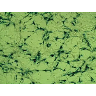
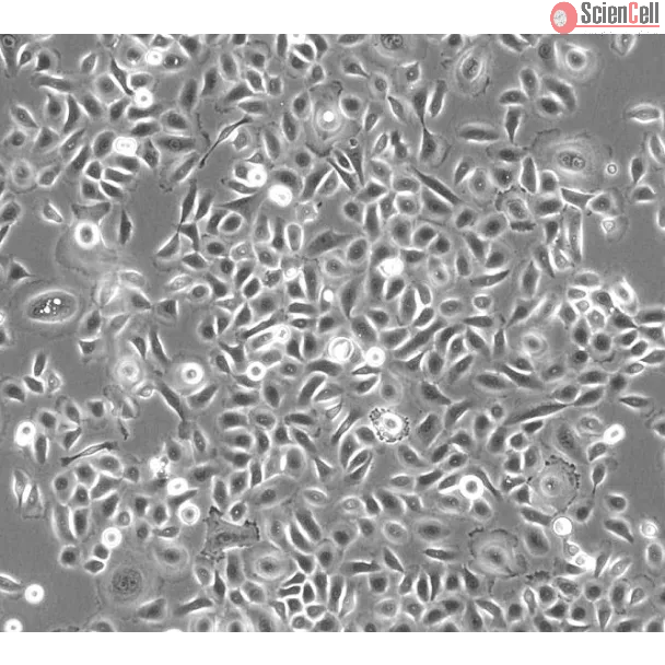
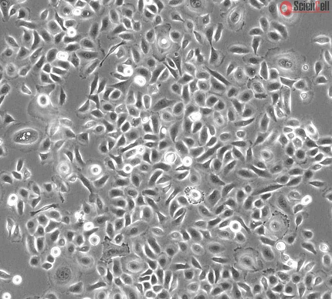
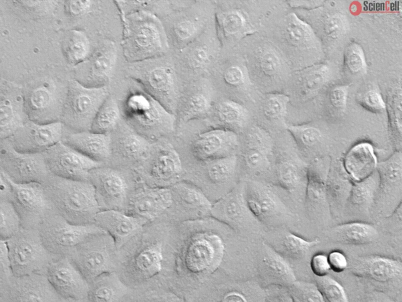
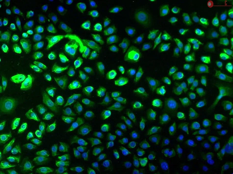
The small airways are located at the interface between alveoli and conducting airways. Airway epithelial cells form a continuous lining in the airways and play a unique role as a protective physical and functional barrier to external deleterious agents. Small airway epithelial cells (SAEpiC) regulate the immune responses by contributing to host defense through chemokine production and adhesion molecule expression. They also produce liquids contributing to pulmonary fluid balance. Many airway diseases, such as asthma, bronchiolitis, chronic obstructive pulmonary disease, and cystic fibrosis, involve damage to the airway surface epithelium. The study of human SAEpiC may help to identify new therapeutic options for preventing airway disorders.
HPSAEpiC from ScienCell Research Laboratories are isolated from human lung tissue. HPSAEpiC are cryopreserved at passage one and delivered frozen. Each vial contains >5 x 10⁵ cells in 1 ml volume. HPSAEpiC are characterized by immunofluorescence with antibodies specific to cytokeratin-18 and/or cytokeratin-19. HPSAEpiC are negative for HIV-1, HBV, HCV, mycoplasma, bacteria, yeast and fungi. HPSAEpiC are guaranteed to further expand for 15 population doublings under the conditions provided by ScienCell Research Laboratories.
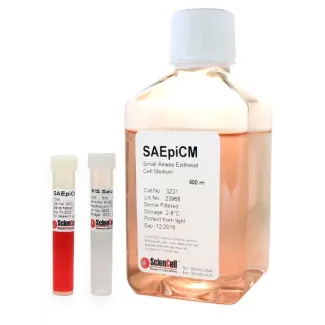
Recommended Medium
It is recommended to use Small Airway Epithelial Cell Medium (SAEpiCM, Cat. #3231) for culturing HPSAEpiC in vitro.
The emergence of diseases associated with telomere dysfunction, including AIDS, aplastic anemia and pulmonary fibrosis, has bolstered interest in telomerase activators. W... More
The emergence of diseases associated with telomere dysfunction, including AIDS, aplastic anemia and pulmonary fibrosis, has bolstered interest in telomerase activators. We report identification of a new small molecule activator, GRN510, with activity ex vivo and in vivo. Using a novel mouse model, we tested the potential of GRN510 to limit fibrosis induced by bleomycin in mTERT heterozygous mice. Treatment with GRN510 at 10 mg/kg/day activated telomerase 2-4 fold both in hematopoietic progenitors ex vivo and in bone marrow and lung tissue in vivo, respectively. Telomerase activation was countered by co-treatment with Imetelstat (GRN163L), a potent telomerase inhibitor. In this model of bleomycin-induced fibrosis, treatment with GRN510 suppressed the development of fibrosis and accumulation of senescent cells in the lung via a mechanism dependent upon telomerase activation. Treatment of small airway epithelial cells (SAEC) or lung fibroblasts ex vivo with GRN510 revealed telomerase activating and replicative lifespan promoting effects only in the SAEC, suggesting that the mechanism accounting for the protective effects of GRN510 against induced lung fibrosis involves specific types of lung cells. Together, these results support the use of small molecule activators of telomerase in therapies to treat idiopathic pulmonary fibrosis. Less
Background: Epithelial-mesenchymal transition (EMT) plays a crucial role in small airway fibrosis of patients with chronic obstructive pulmonary disease (COPD). Increasin... More
Background: Epithelial-mesenchymal transition (EMT) plays a crucial role in small airway fibrosis of patients with chronic obstructive pulmonary disease (COPD). Increasing evidence suggests that the urokinase plasminogen activator receptor (uPAR) is involved in the pathogenesis of COPD. Increased uPAR expression has been implicated in the promotion of EMT in numerous cancers; however the role of uPAR in EMT in small airway epithelial cells of patients with COPD remains unclear. In this study, we investigated the degree of EMT and uPAR expression in lung epithelium of COPD patients, and verified the effect of uPAR on cigarette smoke extract (CSE)-induced EMT in vitro. Methods: The expression of EMT biomarkers and uPAR was assessed in lung epithelium specimens from non-smokers (n = 25), smokers (n = 25) and non-smokers with COPD (n = 10) and smokers with COPD (n = 18). The role of uPAR on CSE-induced EMT in human small airway epithelial cells (HSAEpiCs) was assessed by silencing uPAR expression in vitro. Results: Markers of active EMT and uPAR expression were significantly increased in the small airway epithelium of patients with COPD compared with controls. We also observed a significant correlation between uPAR and vimentin expression in the small airway epithelium. In vitro, CSE-induced EMT in HSAEpiCs was associated with high expression of uPAR, and targeted silencing of uPAR using shRNA inhibited CSE-induced EMT. Finally, we demonstrate that the PI3K/Akt signaling pathway is required for uPAR-mediated EMT in HSAEpiCs. Conclusions: A uPAR-dependent signaling pathway is required for CSE-induced EMT, which contributes to small airway fibrosis in COPD. We propose that increased uPAR expression in the small airway epithelium of patients with COPD participates in an active EMT process. Less
Background and purpose. Metal ion toxicity both locally and systemically following MoM hip replacements remains a concern. Cobalt ions have been shown to induce secretion... More
Background and purpose. Metal ion toxicity both locally and systemically following MoM hip replacements remains a concern. Cobalt ions have been shown to induce secretion of proinflammatory chemokines locally; however, little is known about their effect systemically. We investigated the in vitro effect of cobalt ions on a variety of cell lines by measuring production of the proinflammatory chemokines IL-8 and MCP-1. Less
ScienCell Research Laboratories (SRL) takes pride in being a resource for researchers all over the world. The publications listed here are not meant as an endorsement or confirmation of the reliability of the products.

,-1-mg-ml--2.jpg)