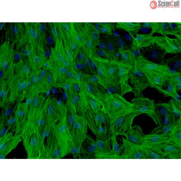
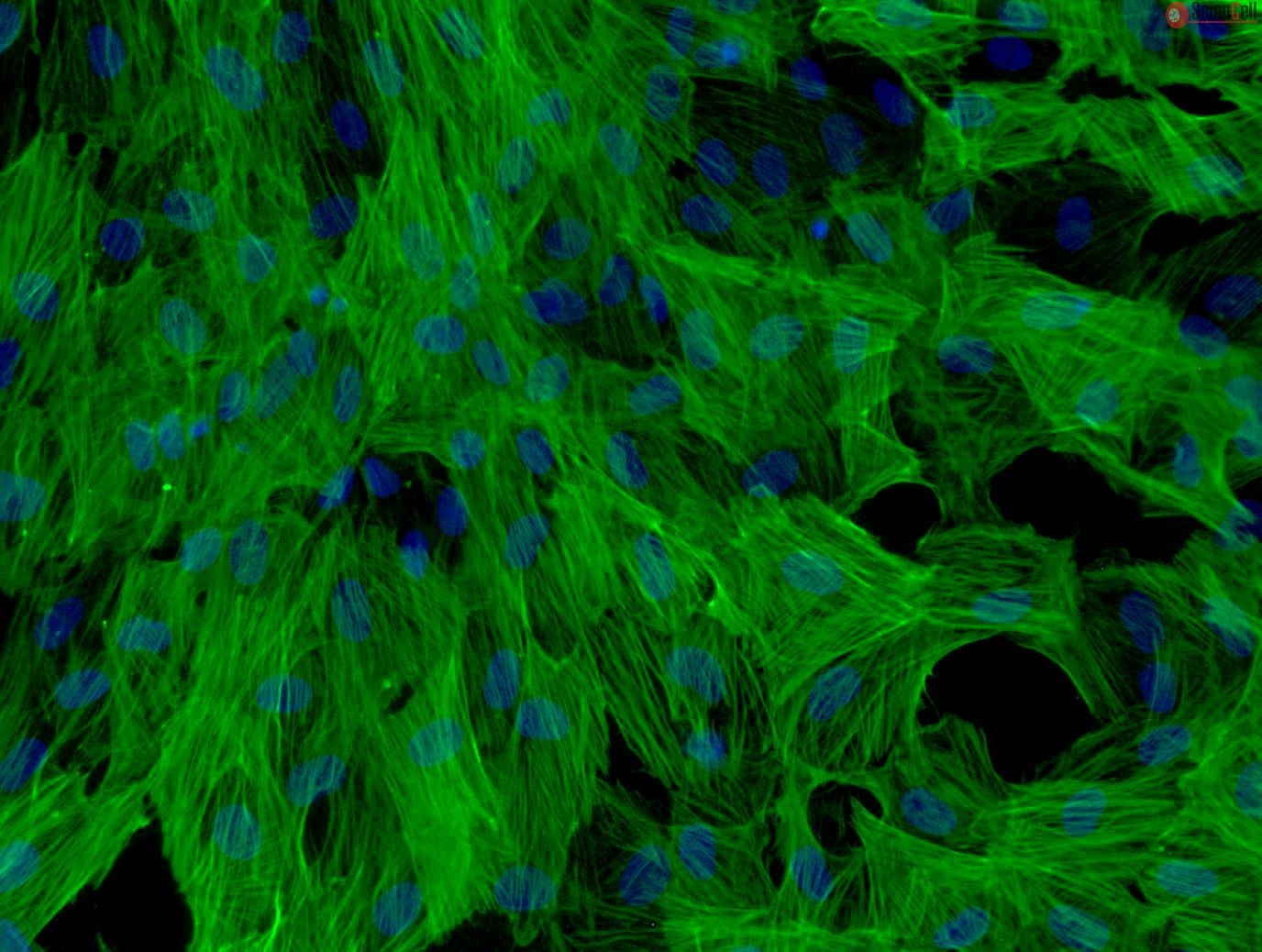
Group 1 innate lymphoid cells (ILCs) comprise a heterogeneous family of cytotoxic natural killer (NK) cells and ILC1s. We identify a population of “liver-type” ILC1s ... More
Group 1 innate lymphoid cells (ILCs) comprise a heterogeneous family of cytotoxic natural killer (NK) cells and ILC1s. We identify a population of “liver-type” ILC1s with transcriptional, phenotypic, and functional features distinct from those of conventional and liver-resident NK cells as well as from other previously described human ILC1 subsets. LT-ILC1s are CD49a+CD94+CD200R1+, express the transcription factor T-BET, and do not express the activating receptor NKp80 or the transcription factor EOMES. Similar to NK cells, liver-type ILC1s produce IFN-γ, TNF-α, and GM-CSF; however, liver-type ILC1s also produce IL-2 and lack perforin and granzyme-B. Liver-type ILC1s are expanded in cirrhotic liver tissues, and they can be produced from blood-derived ILC precursors in vitro in the presence of TGF-β1 and liver sinusoidal endothelial cells. Cells with similar signature and function can also be found in tonsil and intestinal tissues. Collectively, our study identifies and classifies a population of human cross-tissue ILC1s. Less
The innate immune system is essential for controlling viral infection. Hepatitis B virus (HBV) persistently infects human hepatocytes and causes hepatocellular carcinoma.... More
The innate immune system is essential for controlling viral infection. Hepatitis B virus (HBV) persistently infects human hepatocytes and causes hepatocellular carcinoma. However, the innate immune response to HBV infection in vivo remains unclear. Using a tree shrew animal model, we showed that HBV infection induced hepatic interferon (IFN)-γ expression during early infection. Our in vitro study demonstrated that hepatic NK cells produced IFN-γ in response to HBV only in the presence of hepatic F4/80(+) cells. Moreover, extracellular vesicles (EVs) released from HBV-infected hepatocytes contained viral nucleic acids and induced NKG2D ligand expression in macrophages by stimulating MyD88, TICAM-1, and MAVS-dependent pathways. In addition, depletion of exosomes from EVs markedly reduced NKG2D ligand expression, suggesting the importance of exosomes for NK cell activation. In contrast, infection of hepatocytes with HBV increased immunoregulatory microRNA levels in EVs and exosomes, which were transferred to macrophages, thereby suppressing IL-12p35 mRNA expression in macrophages to counteract the host innate immune response. IFN-γ increased the hepatic expression of DDX60 and augmented the DDX60-dependent degradation of cytoplasmic HBV RNA. Our results elucidated the crucial role of exosomes in antiviral innate immune response against HBV.
Accession number: Accession number of RNA-seq data is DRA004164 (DRA in DDBJ).
Keywords: exosome; innate immunity; virus. Less
Background
Immuno-genetic studies suggest a functional link between NK cells and λ-IFNs. We recently showed that NK cells are negative for the IFN-λ receptor IFN-λR1 a... More
Background
Immuno-genetic studies suggest a functional link between NK cells and λ-IFNs. We recently showed that NK cells are negative for the IFN-λ receptor IFN-λR1 and do not respond to IFN-λ, suggesting a rather indirect association between IL-28B genotype and NK cell activity.
Methods
A total of 75 HCV(+) patients and 67 healthy controls were enrolled into this study. IL-28B (rs12979860) and IFNL-4 (rs368234815) genotypes were determined by rtPCR. Total PBMC, monocytes, and NK cells were stimulated with IL-29, the TLR-7/8 agonist R848, or a combination of both. NK cell IFN-γ response was analysed by FACS. IL-12 and IL-18 secretion of monocytes was studied by ELISA. In blocking experiments anti-IL-12/anti-IL-18 were used.
Results
Following stimulation of total PBMCs with R848 we found NK cell IFN- γ responses to vary with the IL-28B genotype, with carriers of a T/T genotype displaying the lowest frequency of IFN-γ(+)NK cells. When isolated NK cells were studied no such associations were observed, indicating an indirect association between IL-28B genotype and NK cell activity.Accordingly, we found R848-stimulated monocytes of patients with a T/T genotype to be significantly less effective in triggering NK cell IFN- γ production than monocytes from carriers of a non-T/T genotype. In line with these findings we observed monocytes from T/T patients to secrete significantly lower concentrations of IL-12 than monocytes from non-T/T individuals.
Conclusions
Our data indicate that monocytes from carriers of an IL-28B T/T genotype display a reduced ability to stimulate NK cell activity and, thus, provide a link between IL-28B genotype and NK functions. Less
Modeling clinically relevant tissue responses using cell models poses a significant challenge for drug development, in particular for drug induced liver injury (DILI). Th... More
Modeling clinically relevant tissue responses using cell models poses a significant challenge for drug development, in particular for drug induced liver injury (DILI). This is mainly because existing liver models lack longevity and tissue-level complexity which limits their utility in predictive toxicology. In this study, we established and characterized novel bioprinted human liver tissue mimetics comprised of patient-derived hepatocytes and non-parenchymal cells in a defined architecture. Scaffold-free assembly of different cell types in an in vivo-relevant architecture allowed for histologic analysis that revealed distinct intercellular hepatocyte junctions, CD31+ endothelial networks, and desmin positive, smooth muscle actin negative quiescent stellates. Unlike what was seen in 2D hepatocyte cultures, the tissues maintained levels of ATP, Albumin as well as expression and drug-induced enzyme activity of Cytochrome P450s over 4 weeks in culture. To assess the ability of the 3D liver cultures to model tissue-level DILI, dose responses of Trovafloxacin, a drug whose hepatotoxic potential could not be assessed by standard pre-clinical models, were compared to the structurally related non-toxic drug Levofloxacin. Trovafloxacin induced significant, dose-dependent toxicity at clinically relevant doses (≤ 4uM). Interestingly, Trovafloxacin toxicity was observed without lipopolysaccharide stimulation and in the absence of resident macrophages in contrast to earlier reports. Together, these results demonstrate that 3D bioprinted liver tissues can both effectively model DILI and distinguish between highly related compounds with differential profile. Thus, the combination of patient-derived primary cells with bioprinting technology here for the first time demonstrates superior performance in terms of mimicking human drug response in a known target organ at the tissue level. Less
Background Hepatic fibrosis is the underlying cause of cirrhosis and liver failure in nearly every form of chronic liver disease, and hepatic stellate cells (HSCs) are th... More
Background Hepatic fibrosis is the underlying cause of cirrhosis and liver failure in nearly every form of chronic liver disease, and hepatic stellate cells (HSCs) are the primary cell type responsible for fibrosis. Long noncoding RNAs (lncRNAs) are increasingly recognized as regulators of development and disease; however, little is known about their expression in human HSCs and their function in hepatic fibrosis. Methods We performed RNA sequencing and ab initio assembly of RNA transcripts to define the lncRNAs expressed in human HSC myofibroblasts. We analyzed chromatin immunoprecipitation data and expression data to identify lncRNAs that were regulated by transforming growth factor beta (TGF-β) signaling, associated with super-enhancers and restricted in expression to HSCs compared with 43 human tissues and cell types. Co-expression network analyses were performed to discover functional modules of lncRNAs, and principle component analysis and K-mean clustering were used to compare lncRNA expression in HSCs with other myofibroblast cell types. Results We identified over 3600 lncRNAs that are expressed in human HSC myofibroblasts. Many are regulated by TGF-β, a major fibrotic signal, and form networks with genes encoding key components of the extracellular matrix (ECM), which is the substrate of the fibrotic scar. The lncRNAs directly regulated by TGF-β signaling are also enriched at super-enhancers. More than 400 of the lncRNAs identified in HSCs are uniquely expressed in HSCs compared with 43 other human tissues and cell types and HSC myofibroblasts demonstrate different patterns of lncRNA expression compared with myofibroblasts originating from other tissues. Co-expression analyses identified a subset of lncRNAs that are tightly linked to collagen genes and numerous proteins that regulate the ECM during formation of the fibrotic scar. Finally, we identified lncRNAs that are induced during progression of human liver disease. Conclusions lncRNAs are likely key contributors to the formation and progression of fibrosis in human liver disease. Less
MicroRNAs (miRNAs) control gene expression by reducing mRNA stability and translation. We aimed to identify alterations in human liver miRNA expression/function in nonalc... More
MicroRNAs (miRNAs) control gene expression by reducing mRNA stability and translation. We aimed to identify alterations in human liver miRNA expression/function in nonalcoholic fatty liver disease (NAFLD). Subjects with the highest (median liver fat 30%, n = 15) and lowest (0%, n = 15) liver fat content were selected from >100 obese patients for miRNA profiling of liver biopsies on microarrays carrying probes for 1438 human miRNAs (a cross-sectional study). Target mRNAs and pathways were predicted for the miRNAs most significantly upregulated in NAFLD, their cell-type-specific expression was investigated by quantitative PCR (qPCR), and the transcriptome of immortalized human hepatocytes (IHH) transfected with the miRNA with the highest number of predicted targets, miR-576-5p, was studied. The screen revealed 42 miRNAs up- and two downregulated in the NAFLD as compared to non-NAFLD liver. The miRNAs differing most significantly between the groups, miR-103a-2*, miR-106b, miR-576-5p, miRPlus-I137*, miR-892a, miR-1282, miR-3663-5p, and miR-3924, were all upregulated in NAFLD liver. Target pathways predicted for these miRNAs included ones involved in cancer, metabolic regulation, insulin signaling, and inflammation. Consistent transcriptome changes were observed in IHH transfected with miR-576-5p, and western analysis revealed a marked reduction of the RAC1 protein belonging to several miR-576-5p target pathways. To conclude, we identified 44 miRNAs differentially expressed in NAFLD versus non-NAFLD liver, 42 of these being novel in the context of NAFLD. The study demonstrates that by applying a novel study set-up and a broad-coverage array platform one can reveal a wealth of previously undiscovered miRNA dysregulation in metabolic disease. Keywords: Hepatocyte; immortalized human hepatocytes; liver biopsy; microRNA; microarray; nonalcoholic fatty liver disease. Less
Background The induction of apoptosis in hepatic stellate cells (HSCs) is a promising therapeutic strategy against hepatitis B virus (HBV)-related hepatic fibrosis. The u... More
Background The induction of apoptosis in hepatic stellate cells (HSCs) is a promising therapeutic strategy against hepatitis B virus (HBV)-related hepatic fibrosis. The underlying mechanisms of apoptosis in HSCs, however, are unknown under consideration of HBV infection. In this study, the effects of HBV on apoptosis and endoplasmic reticulum (ER) stress signaling in HSCs were examined. Methods The effects of conditioned media (CM) from HepG2.2.15 on apoptosis induced by the proteasome inhibitor MG132 in LX-2 and HHSteC were studied in regard to c-Jun. In combination with c-Fos, c-Jun forms the AP-1 early response transcription factor, leading to AP-1 activation, signal transduction, endoplasmic reticulum (ER) stress and apoptosis. Results In LX-2 cells, MG132 treatment was associated with the phosphorylation of c-Jun, activation of AP-1 and apoptosis. However, in the presence of CM from HepG2.2.15, these phenomena were attenuated. In HHSteC cells, similar results were observed. HBV genomic DNA is not involved in the process of HSC apoptosis. It is possible that HBeAg has an inhibitory effect on MG132-induced apoptosis in LX-2. We also observed the upregulation of several ER stress-associated genes, such as cAMP responsive element binding protein 3-like 3, inhibin-beta A and solute carrier family 17-member 2, in the presence of CM from HepG2.2.15, or CM from PXB cells infected with HBV. Conclusions HBV inhibits the activation of c-Jun/AP-1 in HSCs, contributing to the attenuation of apoptosis and resulting in hepatic fibrosis. HBV also up-regulated several ER stress genes associated with cell growth and fibrosis. These mechanistic insights might shed new light on a treatment strategy for HBV-associated hepatic fibrosis. Less
MicroRNA-122 (miR-122) is one of the most abundant miRs in the liver. Previous studies have demonstrated that miR-122 plays a role in inflammation in the liver and functi... More
MicroRNA-122 (miR-122) is one of the most abundant miRs in the liver. Previous studies have demonstrated that miR-122 plays a role in inflammation in the liver and functions in hepatic stellate cells (HSCs), which reside in the space of Disse. Here, we showed that the transient inhibition of PKR-activating protein (PACT) expression, by miR-122 or siRNA targeting of PACT, suppressed the production of proinflammatory cytokines, such as interleukin (IL)-6, monocyte chemoattractant protein-1 (MCP-1) and IL-1β, in human HSC LX-2. Sequence and functional analyses confirmed that miR-122 directly targeted the 3′-untranslated region of PACT. Immunofluorescence analysis revealed that miR-122 blocked NF-κB-nuclear translocation in LX-2 cells. We also showed that conditioned medium from miR-122-transfected LX-2 cells suppressed human monocyte-derived THP-1 cell migration. Taken together, our study indicates that miR-122 may downregulate cytokine production in HSCs and macrophage chemotaxis and that the targeting of miR-122 may have therapeutic potential for preventing the progression of liver diseases. Less
Autophagy, a type II programmed cell death, is essential for cell survival under stress, e.g. lung injury, and bone marrow-derived mesenchymal stem cells (BM-MSCs) have g... More
Autophagy, a type II programmed cell death, is essential for cell survival under stress, e.g. lung injury, and bone marrow-derived mesenchymal stem cells (BM-MSCs) have great potential for cell therapy. However, the mechanisms underlying the BM-MSC activation of autophagy to provide a therapeutic effect in ischaemia/reperfusion-induced lung injury (IRI) remain unclear. Thus, we investigate the activation of autophagy in IRI following transplantation with BM-MSCs. Seventy mice were pre-treated with BM-MSCs before they underwent lung IRI surgery in vivo. Human pulmonary micro-vascular endothelial cells (HPMVECs) were pre-conditioned with BM-MSCs by oxygen-glucose deprivation/reoxygenation (OGD) in vitro. Expression markers for autophagy and the phosphoinositide 3-kinase/protein kinase B (PI3K/Akt) signalling pathway were analysed. In IRI-treated mice, administration of BM-MSCs significantly attenuated lung injury and inflammation, and increased the level of autophagy. In OGD-treated HPMVECs, co-culture with BM-MSCs attenuated endothelial permeability by decreasing the level of cell death and enhanced autophagic activation. Moreover, administration of BM-MSCs decreased the level of PI3K class I and p-Akt while the expression of PI3K class III was increased. Finally, BM-MSCs-induced autophagic activity was prevented using the inhibitor LY294002. Administration of BM-MSCs attenuated lung injury by improving the autophagy level via the PI3K/Akt signalling pathway. These findings provide further understanding of the mechanisms related to BM-MSCs and will help to develop new cell-based therapeutic strategies in lung injury. Less
Current in vitro and animal tests for drug development are failing to emulate the systemic organ complexity of the human body and, therefore, to accurately predict drug t... More
Current in vitro and animal tests for drug development are failing to emulate the systemic organ complexity of the human body and, therefore, to accurately predict drug toxicity. In this study, we present a multi-organ-chip capable of maintaining 3D tissues derived from cell lines, primary cells and biopsies of various human organs. We designed a multi-organ-chip with co-cultures of human artificial liver microtissues and skin biopsies, each a 1/100 000 of the biomass of their original human organ counterparts, and have successfully proven its long-term performance. The system supports two different culture modes: i) tissue exposed to the fluid flow, or ii) tissue shielded from the underlying fluid flow by standard Transwell® cultures. Crosstalk between the two tissues was observed in 14-day co-cultures exposed to fluid flow. Applying the same culture mode, liver microtissues showed sensitivity at different molecular levels to the toxic substance troglitazone during a 6-day exposure. Finally, an astonishingly stable long-term performance of the Transwell®-based co-cultures could be observed over a 28-day period. This mode facilitates exposure of skin at the air–liquid interface. Thus, we provide here a potential new tool for systemic substance testing. Less
To examine the possible involvement of nonparenchymal cells in the development of preneoplastic hepatic lesions induced by clofibrate (CF), alterations of these cells wer... More
To examine the possible involvement of nonparenchymal cells in the development of preneoplastic hepatic lesions induced by clofibrate (CF), alterations of these cells were investigated immunohistochemically in glutathione S-transferase M1 gene polymorphic rats (KS and NC types) with different cancer susceptibilities. After CF administration for 8 weeks, α-smooth muscle actin (α-SMA)-positive hepatic stellate cells (HSC) were markedly decreased in sensitive KS-type rats, but not in the NC-type rats. Kupffer cells were decreased with similar extents between them. The sinusoidal endothelial cells were not changed in either type. The other markers for HSC, vimentin and CRBP1, also confirmed the decrease of HSC in the KS type. The decrease of HSC was not observed at 4 weeks of CF administration. Preneoplastic peroxisomal bifunctional enzyme-negative foci were detected in the KS-type rats at 8 weeks of CF administration, but not at 4 weeks. Human HSC were cultured in the presence of clofibric acid and expression of most HSC marker genes, such as vimentin and α-SMA (ACTA2), evaluated by a microarray, was not altered by the treatment, suggesting that HSC loss in the KS-type rats was not due to the direct toxic effect of CF. The expression levels of most HSC marker genes were low in both control and CF-treated rat livers. A possible link between HSC loss and the development of preneoplastic hepatic foci is discussed. Less
Chemotaxis signals between hepatic stellate cells (HSC) and sinusoidal endothelial cells (SEC) maintain hepatic vascular homeostasis and integrity and also regulate chang... More
Chemotaxis signals between hepatic stellate cells (HSC) and sinusoidal endothelial cells (SEC) maintain hepatic vascular homeostasis and integrity and also regulate changes in sinusoidal structure in response to liver injury. Our prior studies have demonstrated that the bidirectional chemotactic signaling molecules EphrinB2 and EphB4 are expressed in HSC. The aim of our present study was to explore whether and how the EphrinB2/EphB4 system in HSC could promote SEC recruitment, which is essential for sinusoidal structure and remodeling. Stimulation of human HSC (hHSC) with chimeric agonists (2 microg/ml) of either EphrinB2 or EphB4 (EphrinB2 Fc or EphB4 Fc, respectively) significantly increased VEGF mRNA levels in hHSC as assessed by quantitative PCR, with respective small interfering RNAs for EphrinB2 and EphB4 inhibiting this increase (P < 0.05, n = 3). EphrinB2 agonist-induced increase in VEGF mRNA levels in hHSC was associated with increased phosphorylation of Erk and was significantly blocked by U0126 (20 microM), an inhibitor of MEK, which is a kinase upstream from Erk (P < 0.05, n = 3). The EphB4 agonist also significantly increased human VEGF promoter activity (P < 0.05, n = 3) as assessed by promoter reporter luciferase assay in transfected LX2-HSC. This was associated with upregulation of the vasculoprotective transcription factor, Kruppel-like factor 2 (KLF2). In Boyden chamber assays, conditioned media from hHSC stimulated with agonists of EphrinB2 or EphB4 increased SEC chemotaxis in a VEGF-dependent manner, compared with control groups that included basal media with agonists of EphrinB2, EphB4, or HSC-conditioned media from HSC in absence of agonist stimulation (P < 0.05, n = 3). EphB4 expression was detected in situ within liver sinusoidal vessels of rats after carbon tetrachloride-induced liver injury. In summary, activation of the EphrinB2/EphB4 signaling pathway in HSC promotes chemotaxis of SEC through a pathway that involves Erk, KLF2, and VEGF. These studies identify EphrinB2 or EphB4 as a key intermediary that links HSC signal transduction pathways with angiogenesis and sinusoidal remodeling. Less
Nonalcoholic steatohepatitis (NASH), the most common cause of chronic liver fibrosis, progresses to cirrhosis in up to 20% of patients. We report that hepatic stellate ce... More
Nonalcoholic steatohepatitis (NASH), the most common cause of chronic liver fibrosis, progresses to cirrhosis in up to 20% of patients. We report that hepatic stellate cells (HSC) in sinusoidal lesions of liver of patients with NASH express high levels of high-affinity IL-13R (IL-13Ralpha2), which is colocalized with smooth muscle actin, whereas fatty liver and normal liver specimens do not express IL-13Ralpha2. HSCs engineered to overexpress IL-13Ralpha2 respond to IL-13 and induce TGFB1 promoter activity and TGF-beta1 production. We also developed NASH in rats by feeding a choline-deficient l-amino acid diet. These rats developed liver fibrosis as assessed by H&E staining, Masson's trichrome and Sirius red staining, and hydroxyproline assays. Treatment of these rats with IL-13R-directed cytotoxin caused a substantial decline in fibrosis and liver enzymes without organ toxicity. These studies demonstrate that functional IL-13Ralpha2 are overexpressed in activated HSCs involved in NASH and that IL-13 cytotoxin ameliorates pathological features of NASH in rat liver, indicating a novel role of this cytotoxin in potential therapy. Less
Background & aims: Cirrhosis is associated with prominent changes in sinusoidal structure and function. Although the resident pericyte in liver, the hepatic stellate cell... More
Background & aims: Cirrhosis is associated with prominent changes in sinusoidal structure and function. Although the resident pericyte in liver, the hepatic stellate cell (HSC), is well characterized in the process of fibrogenesis, signaling pathways that regulate HSC vascular function are less developed. Because pericyte populations outside the liver are increasingly being recognized as a key cell type for angiogenesis and changes in vascular structure, in this study, we explore new HSC-signaling pathways that regulate sinusoidal structure and function. Methods: Real-time video microscopy and quantitative software analysis of vascular tube formation were used to measure HSC angiogenesis in vitro. Platelet-derived growth factor (PDGF) and ephrin-signaling pathways were modulated using molecular and pharmacologic techniques. Complementary whole animal studies were performed to correlate in vitro findings with pericyte functions in vivo. Results: We show that PDGF promotes a phenotype of HSC evidenced by enhanced HSC-driven vascular tube formation in vitro and enhanced HSC coverage of sinusoids in vivo. This angiogenic phenotype modulates specific pericyte vascular functions including permeability and pressure regulation. Furthermore, we identify a key role for ephrin-B2 as a downstream effector of PDGF signaling. Conclusions: These studies elucidate novel HSC-signaling pathways that regulate microvascular structure and function in liver. Less
Chronic hepatitis B virus (HBV) infection is a major cause of liver fibrosis, eventually leading to cirrhosis and hepatocellular carcinoma. Although the involvement of th... More
Chronic hepatitis B virus (HBV) infection is a major cause of liver fibrosis, eventually leading to cirrhosis and hepatocellular carcinoma. Although the involvement of the X protein of HBV (HBx) in viral replication and tumor development has been extensively studied, little is known about its possible role in the development of fibrosis. In this work we show that expression of HBx in hepatocytes results in paracrine activation and proliferation of hepatic stellate cells (HSCs), the main producers of extracellular matrix proteins in the fibrotic liver. Both human primary HSCs and rat HSCs exposed to conditioned medium from HBx-expressing hepatocytes showed increased expression of collagen I, connective tissue growth factor, alpha smooth muscle actin, matrix metalloproteinase-2, and transforming growth factor-beta (TGF-beta), together with an enhanced proliferation rate. We found that HBx induced TGF-beta secretion in hepatocytes and that the activation of HSCs by conditioned medium from HBx-expressing hepatocytes was prevented by a neutralizing anti-TGF-beta antibody, indicating the involvement of this profibrotic factor in the process. Conclusion: Our results propose a direct role for HBx in the development of liver fibrosis by the paracrine activation of stellate cells and reinforce the indication of antiviral treatment in patients with advanced HBV-related chronic liver disease and persistent liver replication. Less
Background/aims: Liver injury results in the activation of hepatic stellate cells (HSCs), which in turn produce matrix metalloproteinase (MMP) in response to pro-inflamma... More
Background/aims: Liver injury results in the activation of hepatic stellate cells (HSCs), which in turn produce matrix metalloproteinase (MMP) in response to pro-inflammatory cytokines for tissue remodelling. This study explored the transcriptional induction of the MMP-1 gene by tumour necrosis factor-alpha (TNF-alpha) in HSCs. Methods: The LI90 human HSC line was used in the present study. Gelatin zymography, enzyme-linked immunosorbent assay, Northern blotting and gene promoter-reporter assays were used to analyse the induction of MMP-1 protein, mRNA expression and gene transcription respectively. Deletional or site-directed mutations were introduced into the promoter region and transiently transfected into LI90 cells to determine the cis-acting elements necessary for TNF-alpha inducibility. Gel shift mobility assays were used to determine the transcriptional factors involved in the TNF-alpha responsiveness. Results: TNF-alpha upregulated MMP-1 protein and mRNA expression in a dose-dependent manner. A time-course experiment revealed a rapid induction of MMP-1 mRNA expression after TNF-alpha treatment. Mutation in a putative nuclear factor (NF)-kappaB-binding site at -2541 bp almost completely abolished the TNF-alpha response to MMP-1 gene-promoter activity, suggesting transcriptional regulation of MMP-1 expression by TNF-alpha via this site. Electrophoretic mobility shift assay and supershift assays indicated that this transcriptional regulation was regulated via the p50/p50 homodimer of NF-kappaB. Conclusions: MMP-1 gene expression might be induced by TNF-alpha via the p50/p50 homodimer of NF-kappaB in activated human HSCs. Less
Prior studies indicate that adenosine and the adenosine A2A receptor play a role in hepatic fibrosis by a mechanism that has been proposed to involve direct stimulation o... More
Prior studies indicate that adenosine and the adenosine A2A receptor play a role in hepatic fibrosis by a mechanism that has been proposed to involve direct stimulation of hepatic stellate cells (HSCs). The objective of this study was to determine whether primary hepatic stellate cells produce collagen in response to adenosine (via activation of adenosine A2A receptors) and to further determine the signaling mechanisms involved in adenosine A2A receptor-mediated promotion of collagen production. Cultured primary HSCs increase their collagen production after stimulation of the adenosine A2A receptor in a dose-dependent fashion. Likewise, LX-2 cells, a human HSC line, increases expression of procollagen alphaI and procollagen alphaIII mRNA and their translational proteins, collagen type I and type III, in response to pharmacological stimulation of adenosine A2A receptors. Based on the use of pharmacological inhibitors of signal transduction, adenosine A2A receptor-mediated stimulation of procollagen alphaI mRNA and collagen type I collagen expression were regulated by signal transduction involving protein kinase A, src, and mitogen-activated protein kinase kinase/extracellular signal-regulated kinase (erk), but surprisingly, adenosine A2A receptor-mediated stimulation of procollagen alphaIII mRNA and collagen type III protein expression depend on the activation of p38 mitogen-activated protein kinase (MAPK), findings confirmed by small interfering RNA-mediated knockdown of src, erk1, erk2, and p38 MAPK. These results indicate that adenosine A2A receptors signal for increased collagen production by multiple signaling pathways. These results provide strong evidence in support of the hypothesis that adenosine receptors promote hepatic fibrosis, at least in part, via direct stimulation of collagen expression and that signaling for collagen production proceeds via multiple pathways. Less
NO antagonizes hepatic stellate cell (HSC) contraction, although activated HSC in cirrhosis demonstrate impaired responses to NO. Decreased NO responses in activated HSC ... More
NO antagonizes hepatic stellate cell (HSC) contraction, although activated HSC in cirrhosis demonstrate impaired responses to NO. Decreased NO responses in activated HSC and mechanisms by which NO affects activated HSC remain incompletely understood. In normal rat HSC, the NO donor diethylamine NONOate (DEAN) significantly increased cGMP production and reduced serum-induced contraction by 25%. The guanylate cyclase (sGC) inhibitor 1H-[1,2,4]oxadiazolo-[4,3-a]quinoxalin-1-one (ODQ) abolished 50% of DEAN effects, whereas the cGMP analog 8-bromoguanosine 3',5'-cyclic monophosphate (8-BrcGMP) reiterated half the observed DEAN response, suggesting both cGMP-dependent protein kinase G (PKG)-dependent and -independent mechanisms of NO-mediated antagonism of normal HSC contraction. However, NO donors did not increase cGMP production from in vivo activated HSC from bile duct-ligated rats and showed alterations in intracellular Ca(2+) accumulation suggesting defective cGMP-dependent effector pathways. The LX-2 cell line also demonstrated lack of cGMP generation in response to NO and a lack of effect of ODQ and 8-BrcGMP in modulating the NO response. However, cGMP-independent effects in response to NO were maintained in LX-2 and were associated with S-nitrosylation of proteins, an effect reiterated in primary HSC. Adenovirus-based overexpression of PKG significantly attenuated contraction of LX-2 by 25% in response to 8-BrcGMP. In summary, these studies demonstrate that NO affects HSC through cGMP-dependent and -independent pathways. The HSC activation process is associated with maintenance of cGMP-independent actions of NO but defects in cGMP-PKG-dependent NO signaling that are improved by PKG gene delivery in LX-2 cells. Activating targets downstream from NO-cGMP in activated HSC may represent a novel therapeutic target for portal hypertension. Less
HSP47 is a collagen-specific chaperone that is required for normal collagen synthesis. In animal models of liver injury, hepatic stellate cells (HSC) have been identified... More
HSP47 is a collagen-specific chaperone that is required for normal collagen synthesis. In animal models of liver injury, hepatic stellate cells (HSC) have been identified as a source of HSP47. Because expression of HSP47 has not been investigated in human liver, the aim of these studies was to characterize expression of HSP47 in human liver and to investigate its regulation in human HSC in vitro. Immunohistochemistry demonstrated staining for HSP47 along the sinusoids of normal and cirrhotic human livers and in fibrous septa. Dual fluorescence confocal microscopy showed colocalization of HSP47 with synaptophysin, a marker for HSC. Levels of immunoreactive HSP47 and its transcript tended to be higher in cirrhotic livers than in normal livers. The abundance of HSP47 protein was unchanged by treatment of cultured human HSC with TGF-β1, angiotensin II, hypoxia and a number of other treatments intended to increase collagen synthesis. A modest reduction in HSP47 was achieved by transfection with antisense oligonucleotides and was associated with a significant decrease in procollagen synthesis. These observations suggest that HSP47 is constitutively expressed in human HSC and that HSP47 may be a target for antifibrotic therapy. Less


,-1-mg-ml--2.jpg)