Fibroblasts are mesenchymal cells derived from the embryonic mesoderm. They have been extensively used for a wide range of cellular and molecular studies as they are one of the easiest types of cells to grow in culture. Their durability also makes them amenable to a variety of manipulations ranging from studies employing gene transfection to microinjection. In general, fibroblasts secrete a non-rigid extracellular matrix which is rich in type I and/or type III collagen. There is evidence showing that fibroblasts in various organs are intrinsically different. Dermal fibroblasts switch from a proliferative, migratory phase to a contractile, matrix-remodeling phase during wound healing. In addition, they secrete large quantities of hyaluronan in response to inflammatory stimuli.
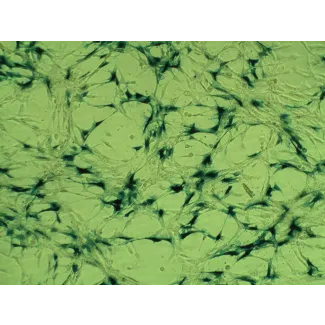
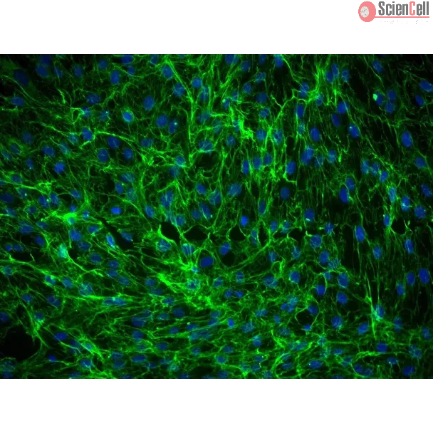
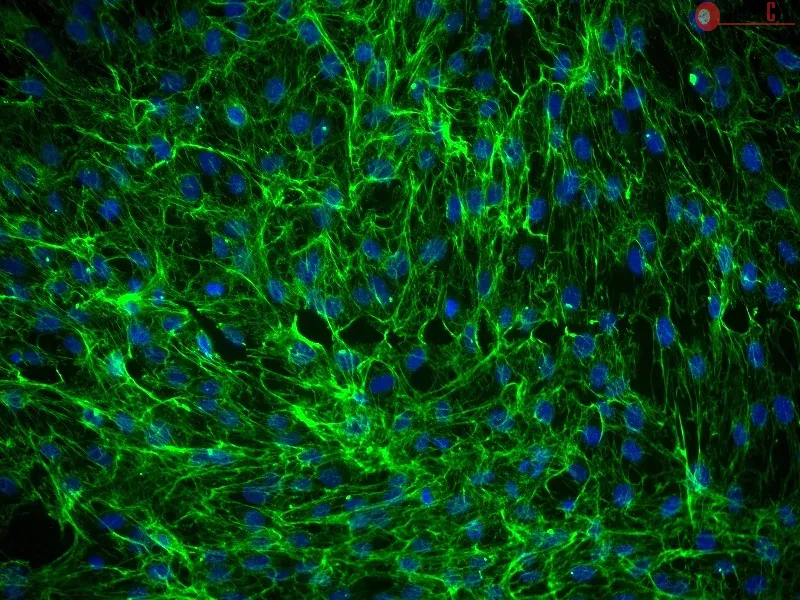
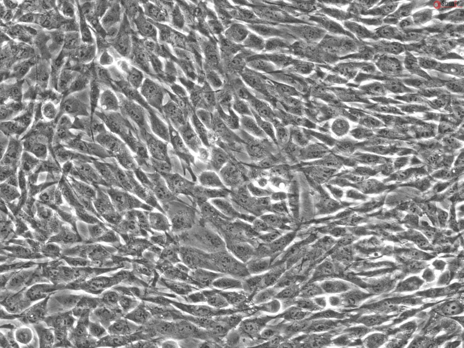
HDF-f from ScienCell Research Laboratories are isolated from fetal human skin. HDF-f are cryopreserved at passage one and delivered frozen. Each vial contains > 5 x 10⁵ cells in 1 ml volume. HDF-f are characterized by their spindle morphology and immunofluorescence with antibody specific to fibronectin. HDF-f are negative for HIV-1, HBV, HCV, mycoplasma, bacteria, yeast, and fungi. HDF-f are guaranteed to further expand for 15 population doublings under the conditions provided by ScienCell Research Laboratories
Recommended Medium
It is recommended to use Fibroblast Medium (FM, Cat. #2301) for culturing HDF-f in vitro.
This study reports on probing the utility of in situ chromatin texture features such as nuclear DNA methylation and chromatin condensation patterns — visualized by fluo... More
This study reports on probing the utility of in situ chromatin texture features such as nuclear DNA methylation and chromatin condensation patterns — visualized by fluorescent staining and evaluated by dedicated three-dimensional (3D) quantitative and high-throughput cell-by-cell image analysis — in assessing the proliferative capacity, i.e. growth behavior of cells: to provide a more dynamic picture of a cell population with potential implications in basic science, cancer diagnostics/prognostics and therapeutic drug development. Two types of primary cells and four different cancer cell lines were propagated and subjected to cell-counting, flow cytometry, confocal imaging, and 3D image analysis at various points in culture. Additionally a subset of primary and cancer cells was accelerated into senescence by oxidative stress. DNA methylation and chromatin condensation levels decreased with declining doubling times when primary cells aged in culture with the lowest levels reached at the stage of proliferative senescence. In comparison, immortal cancer cells with constant but higher doubling times mostly displayed lower and constant levels of the two in situ-derived features. However, stress-induced senescent primary and cancer cells showed similar levels of these features compared with primary cells that had reached natural growth arrest. With regards to global DNA methylation and chromatin condensation levels, aggressively growing cancer cells seem to take an intermediate level between normally proliferating and senescent cells. Thus, normal cells apparently reach cancer-cell equivalent stages of the two parameters at some point in aging, which might challenge phenotypic distinction between these two types of cells. Companion high-resolution molecular profiling could provide information on possible underlying differences that would explain benign versus malign cell growth behaviors. Keywords: DNA methylation, chromatin condensation, cell proliferation, aging, senescence, cancer, 3D imaging, cell-by-cell analysis Less
Reprogramming human somatic cells into pluripotent cells opens up new possibilities for transplantation therapy, the study of disease, and drug screening. In addition to ... More
Reprogramming human somatic cells into pluripotent cells opens up new possibilities for transplantation therapy, the study of disease, and drug screening. In addition to somatic cell nuclear transfer, several approaches to reprogramming human cells have been reported: transduction of defined transcription factors to generate induced pluripotent stem cell (iPSC), human embryonic stem cell (hESC)-somatic cell fusion, and hESC cytoplast-somatic cell fusion or exposure to extracts of hESC. Here, we optimized techniques for hESC-human fibroblast fusion and enucleation and cytoplast fusion, and then compared the reprogramming efficiency between iPSC generation, cell-fusion and cytoplast-fusion. When compared with iPSC, hESC-fusion provided much faster and efficient reprogramming of somatic cells. The reprogramming required more than 4 weeks and the efficiency was less than 0.001% in iPSC generation, and it was less than 10 days and more than 0.005% in hESC-fusion. In addition, fusion yielded almost no partially reprogrammed cell colonies. However, the fused cells were tetraploid or aneuploid. hESC cytoplast fusion could initiate reprogramming but was never able to complete reprogramming. These data indicate that in cell fusion, as in nuclear transfer, reprogramming through direct introduction of a somatic nucleus into the environment of a pluripotent cell provides relatively efficient reprogramming. The findings also suggest that the nucleus of the host pluripotent cell may contain components that accelerate the reprogramming process. Less
Gene delivery into human hepatocytes remains a critical issue for the development of liver-directed gene therapy. Gene delivery based on non-viral vectors is an attractiv... More
Gene delivery into human hepatocytes remains a critical issue for the development of liver-directed gene therapy. Gene delivery based on non-viral vectors is an attractive approach relative to viral vectors. In this report, novel delivery system of preS/liposome/DNA virus-like particle (VLP) was developed for gene transfection into hepatocytes in vivo and in vitro. Plasmid pCMVbeta, expressing beta-galactosidase, was encapsulated with cationic liposome, and then the histidine-tagged preS domain of hepatitis B virus was coated on the surface of liposome/DNA to form preS/liposome/ DNA VLP. Transfection efficiencies of preS/liposome/DNA, liposome/DNA, naked DNA and preS were analyzed using several different human cell lines. The highest transfection efficiency was found using preS/liposome/DNA VLP as the transfection reagent in human hepatocyte (HH) cell line. Results show that preS domain of hepatitis B virus coated on liposome/DNA can be used for highly efficient gene transfection into human hepatocytes. Moreover, the target characteristic of preS/liposome/DNA was analyzed in vivo. After preS/liposome/DNA VLP was injected into immunocompromised (Nude) mice via the tail vein, most of beta-galactosidase was expressed in the liver; however, no significant target expression was found with the injection of liposome/ DNA or naked DNA. Our results show that preS/liposome/DNA VLP can be used as a novel liver-specific gene delivery system. Less
ScienCell Research Laboratories (SRL) takes pride in being a resource for researchers all over the world. The publications listed here are not meant as an endorsement or confirmation of the reliability of the products.


,-1-mg-ml--2.jpg)
.webp)