Human induced pluripotent stem cells (iPS cells) hold great promise in the field of regenerative medicine, especially immune-compatible cell therapy. The most important s... More
Human induced pluripotent stem cells (iPS cells) hold great promise in the field of regenerative medicine, especially immune-compatible cell therapy. The most important safety-related issues that must be resolved before the clinical use of iPS cells include the generation of “footprint-free” and “xeno-free” iPS cells. In this study, we sought to examine whether an extracellular matrix- (ECM-) based xeno-free culture system that we recently established could be used together with a microRNA-enhanced mRNA reprogramming method for the generation of clinically safe iPS cells. The notable features of this method are the use of a xeno-free/feeder-free culture system for the generation and expansion of iPS cells rather than the conventional labor-intensive culture systems using human feeder cells or human feeder-conditioned medium and the enhancement of mRNA-mediated reprogramming via the delivery of microRNAs. Strikingly, we observed the early appearance of iPS cell colonies (~11 days), substantial reprogramming efficiency (~0.2–0.3%), and a high percentage of ESC-like colonies among the total colonies (~87.5%), indicating enhanced kinetics and reprogramming efficiency. Therefore, the combined method established in this study provides a valuable platform for the generation and expansion of clinically safe (i.e., integration- and xeno-free) iPS cells, facilitating immune-matched cell therapy in the near future. Less
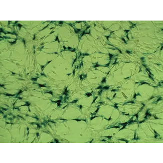
In stem cell cultures from adult human tissue, undesirable contamination with fibroblasts is frequently present. The presence of fibroblasts obscures the actual number of... More
In stem cell cultures from adult human tissue, undesirable contamination with fibroblasts is frequently present. The presence of fibroblasts obscures the actual number of stem cells and may result in extracellular matrix production after transplantation. Identification of fibroblasts is difficult because of the lack of specific fibroblast markers. In our laboratory, we isolate and expand neural-crest-derived stem cells from human hair follicle bulges and investigate their potential to differentiate into neural cells. To establish cellular identities, we perform immunohistochemistry with antibodies specific for glial and neuronal markers, and use fibroblasts as negative control. We frequently observe that human adult dermal fibroblasts also express some glial and neuronal markers. In this study, we have sought to determine whether our observations represent actual expression of these markers or result from cross-reactivity. Immunohistochemistry was performed on human adult dermal fibroblasts using acknowledged glial and neuronal antibodies followed by verification of the data using RT-qPCR. Human adult dermal fibroblasts showed expression of the glia-specific markers SOX9, glial fibrillary acidic protein and EGR2 (KROX20) as well as for the neuron-specific marker class III β-tubulin, both at the protein and mRNA level. Furthermore, human adult dermal fibroblasts showed false-positive immunostaining for S100β and GAP43 and to a lower extent for OCT6. Our results indicate that immunophenotyping as a tool to determine cellular identity is not as reliable as generally assumed, especially since human adult dermal fibroblasts may be mistaken for neural cells, indicating that the ultimate proof of glial or neuronal identity can only be provided by their functionality. Less
Electrical stimulation (ES) has long been used as an alternative clinical treatment and an effective approach to modulate cellular behaviours. In this work we investigate... More
Electrical stimulation (ES) has long been used as an alternative clinical treatment and an effective approach to modulate cellular behaviours. In this work we investigated the effects of ES on human skin fibroblast activity, myofibroblast transdifferentiation and the consequence on wound healing. Normal human fibroblasts were seeded on heparin-bioactivated PPy/PLLA conductive membranes, cultured for 24 h, and then exposed to ES of 50 or 200 mV/mm for 2, 4, or 6 h. Following ES, the cells were either subjected to various analyses or re-seeded to investigate their healing capacity. Our findings show that ES had no cytotoxic effect on the fibroblasts, as demonstrated by the similar LDH activity levels in the ES-exposed and non-exposed cultures, and by the comparable cell viability under both conditions. Furthermore, the number of viable fibroblasts was higher following exposure to 6 h of ES than in the non-exposed culture. This enhanced cell growth was likely due to the ES up-regulated secretion of FGF-1 and FGF-2. In an in vitro scratch-wound assay where cell monolayer was used as a healing model, the electrically stimulated dermal fibroblasts migrated faster following exposure to ES and recorded a high contractile behaviour toward the collagen gel matrix. This enhanced contraction was supported by the high level of α-smooth muscle actin expressed by the fibroblasts following exposure to ES, indicating the characteristics of myofibroblasts. Remarkably, the modulation of fibroblast growth continued long after ES. In conclusion, this work demonstrates for the first time that exposure to ES promoted skin fibroblast growth and migration, increased growth factor secretion, and promoted fibroblast to myofibroblast transdifferentiation, thus promoting wound healing. Less
Background: Macrophages and fibroblasts are two major players in tissue repair and fibrosis. Despite the relevance of macrophages and fibroblasts in tissue homeostasis, r... More
Background: Macrophages and fibroblasts are two major players in tissue repair and fibrosis. Despite the relevance of macrophages and fibroblasts in tissue homeostasis, remarkably little is known whether macrophages are able to influence the properties of fibroblasts. Here we investigated the role of paracrine factors secreted by classically activated (M1) and alternatively activated (M2) human macrophages on human dermal fibroblasts (HDFs). Results: HDFs stimulated with paracrine factors from M1 macrophages showed a 10 to > 100-fold increase in the expression of the inflammatory cytokines IL6, CCL2 and CCL7 and the matrix metalloproteinases MMP1 and MMP3. This indicates that factors produced by M1 macrophages induce a fibroblast phenotype with pro-inflammatory and extracellular matrix (ECM) degrading properties. HDFs stimulated with paracrine factors secreted by M2 macrophages displayed an increased proliferation rate. Interestingly, the M1-activated pro-inflammatory fibroblasts downregulated, after exposure to paracrine factors produced by M2 macrophages or non-conditioned media, the inflammatory markers as well as MMPs and upregulated their collagen production. Conclusions: Paracrine factors of M1 or M2 polarized macrophages induced different phenotypes of HDFs and the HDF phenotypes can in turn be reversed, pointing to a high dynamic plasticity of fibroblasts in the different phases of tissue repair. Less
This study is the first to investigate the anticancer effects of the new phloroglucinol derivative (3,6-bis(3-chlorophenylacetyl)phloroglucinol; MCPP) in human colon canc... More
This study is the first to investigate the anticancer effects of the new phloroglucinol derivative (3,6-bis(3-chlorophenylacetyl)phloroglucinol; MCPP) in human colon cancer cells. MCPP induced cell death and antiproliferation in three human colon cancer, HCT-116, SW480, and Caco-2 cells, but not in primary human dermal fibroblast cells. MCPP-induced concentration-dependent apoptotic cell death in colon cancer cells was measured by fluorescence-activated cell sorter (FACS) analysis. Treatment of HCT-116 human colon cancer cells with MCPP was found to induce a number of signature endoplasmic reticulum (ER) stress markers; and up-regulation of CCAAT/enhancer-binding protein homologous protein (CHOP) and glucose-regulated protein (GRP)-78, phosphorylation of eukaryotic initiation factor-2α (eIF-2α), suggesting the induction of ER stress. MCPP also increased GSK3α/β(Tyr270/216) phosphorylation and reduced GSK3α/β(Ser21/9) phosphorylation time-dependently. Transfection of cells with GRP78 or CHOP siRNA, or treatment of GSK3 inhibitor SB216163 reduced MCPP-mediated cell apoptosis. Treatment of MCPP also increased caspase-7, caspase-9, and caspase-3 activity. The inhibition of caspase activity by z-DEVE-FMK or z-VAD-FMK significantly reduced MCPP-induced apoptosis. Furthermore, treatment of GSK3 inhibitor SB216763 also dramatically reversed MCPP-induced GRP and CHOP up-regulation, and pro-caspase-3 and pro-caspase-9 degradation. Taken together, the present study provides evidences to support that GRP78 and CHOP expression, and GSK3α/β activation in mediating the MCPP-induced human colon cancer cell apoptosis. Copyright © 2010 Wiley-Liss, Inc. Less
We have used microarray-based methods of global gene expression together with quantitative PCR and Western blot analysis to identify dysregulation of genes and aberrant c... More
We have used microarray-based methods of global gene expression together with quantitative PCR and Western blot analysis to identify dysregulation of genes and aberrant cellular processes in human fibroblasts and in SH-SY5Y neuroblastoma cells made HPRT-deficient by transduction with a retrovirus stably expressing an shRNA targeted against HPRT. Analysis of the microarray expression data by Gene ontology (GO) and Gene Set Enrichment Analysis (GSEA) as well as significant pathway analysis by GeneSpring GX10 and Panther Classification System reveal that HPRT deficiency is accompanied by aberrations in a variety of pathways known to regulate neurogenesis or to be implicated in neurodegenerative disease, including the canonical Wnt/β-catenin and the Alzheimer's disease/presenilin signaling pathways. Dysregulation of the Wnt/β-catenin pathway is confirmed by Western blot demonstration of cytosolic sequestration of β-catenin during in vitro differentiation of the SH-SY5Y cells toward the neuronal phenotype. We also demonstrate that two key transcription factor genes known to be regulated by Wnt signaling and to be vital for the generation and function of dopaminergic neurons; i.e., Lmx1a and Engrailed 1, are down-regulated in the HPRT knockdown SH-SY5Y cells. In addition to the Wnt signaling aberration, we found that expression of presenilin-1 shows severely aberrant expression in HPRT-deficient SH-SY5Y cells, reflected by marked deficiency of the 23 kDa C-terminal fragment of presenilin-1 in knockdown cells. Western blot analysis of primary fibroblast cultures from two LND patients also shows dysregulated presenilin-1 expression, including aberrant proteolytic processing of presenilin-1. These demonstrations of dysregulated Wnt signaling and presenilin-1 expression together with impaired expression of dopaminergic transcription factors reveal broad pleitropic neuro-regulatory defects played by HPRT expression and suggest new directions for investigating mechanisms of aberrant neurogenesis and neuropathology in LND and potential new targets for restoration of effective signaling in this neuro-developmental defect. Less
We present a method to create multi-layered engineered tissue composites consisting of human skin fibroblasts and keratinocytes which mimic skin layers. Three-dimensional... More
We present a method to create multi-layered engineered tissue composites consisting of human skin fibroblasts and keratinocytes which mimic skin layers. Three-dimensional (3D) freeform fabrication (FF) technique, based on direct cell dispensing, was implemented using a robotic platform that prints collagen hydrogel precursor, fibroblasts and keratinocytes. A printed layer of cell-containing collagen was crosslinked by coating the layer with nebulized aqueous sodium bicarbonate. The process was repeated in layer-by-layer fashion on a planar tissue culture dish, resulting in two distinct cell layers of inner fibroblasts and outer keratinocytes. In order to demonstrate the ability to print and culture multi-layered cell-hydrogel composites on a non-planar surface for potential applications including skin wound repair, the technique was tested on a poly(dimethylsiloxane) (PDMS) mold with 3D surface contours as a target substrate. Highly viable proliferation of each cell layer was observed on both planar and non-planar surfaces. Our results suggest that organotypic skin tissue culture is feasible using on-demand cell printing technique with future potential application in creating skin grafts tailored for wound shape or artificial tissue assay for disease modeling and drug testing. Less
The inflammatory response plays out over time in a reproducible and organized way after an initiating stimulus. Here we show that genes activated in cultured mouse fibrob... More
The inflammatory response plays out over time in a reproducible and organized way after an initiating stimulus. Here we show that genes activated in cultured mouse fibroblasts in response to the cytokine tumor necrosis factor could be categorized into roughly three groups, each with different induction kinetics. Although differences in transcription were important in determining the grouping of these genes, differences in mRNA stability also exerted a strong influence on the temporal order of gene expression, in some cases overriding that of transcriptional control elements. Transcripts of mRNA expressed early had abundant AU-rich elements in their 3' untranslated regions, whereas those expressed later had fewer. Thus, mRNA stability and transcriptional control, two intrinsic characteristics of genes, control the kinetics of gene expression induced by proinflammatory cytokines. Less
We present a method to create multi-layered engineered tissue composites consisting of human skin fibroblasts and keratinocytes which mimic skin layers. Three-dimensional... More
We present a method to create multi-layered engineered tissue composites consisting of human skin fibroblasts and keratinocytes which mimic skin layers. Three-dimensional (3D) freeform fabrication (FF) technique, based on direct cell dispensing, was implemented using a robotic platform that prints collagen hydrogel precursor, fibroblasts and keratinocytes. A printed layer of cell-containing collagen was crosslinked by coating the layer with nebulized aqueous sodium bicarbonate. The process was repeated in layer-by-layer fashion on a planar tissue culture dish, resulting in two distinct cell layers of inner fibroblasts and outer keratinocytes. In order to demonstrate the ability to print and culture multi-layered cell-hydrogel composites on a non-planar surface for potential applications including skin wound repair, the technique was tested on a poly(dimethylsiloxane) (PDMS) mold with 3D surface contours as a target substrate. Highly viable proliferation of each cell layer was observed on both planar and non-planar surfaces. Our results suggest that organotypic skin tissue culture is feasible using on-demand cell printing technique with future potential application in creating skin grafts tailored for wound shape or artificial tissue assay for disease modeling and drug testing. Less
Senescent and suppressor T cells are reported to be increased in select patients with cancer and are poor prognostic indicators. Based on the association of these T cells... More
Senescent and suppressor T cells are reported to be increased in select patients with cancer and are poor prognostic indicators. Based on the association of these T cells and poor outcomes, we hypothesized that tumors induce senescence in T cells, which negatively effects antitumor immunity. In this report, we show that human T cells from healthy donors incubated with tumor for only 6 h at a low tumor to T-cell ratio undergo a senescence-like phenotype, characterized by the loss of CD27 and CD28 expression and telomere shortening. Tumor-induced senescence of T cells is induced by soluble factors and triggers increases in expression of senescence-associated molecules such as p53, p21, and p16. Importantly, these T cells are not only phenotypically altered, but also functionally altered as they can suppress the proliferation of responder T cells. This suppression requires cell-to-cell contact and is mediated by senescent CD4(+) and CD8(+) subpopulations, which are distinct from classically described natural T regulatory cells. Our observations support the novel concept that tumor can induce senescent T cells with suppressor function and may effect both the diagnosis and treatment of cancer. Less


,-1-mg-ml--2.jpg)