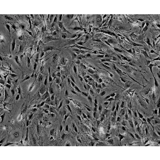
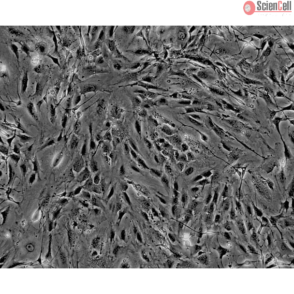
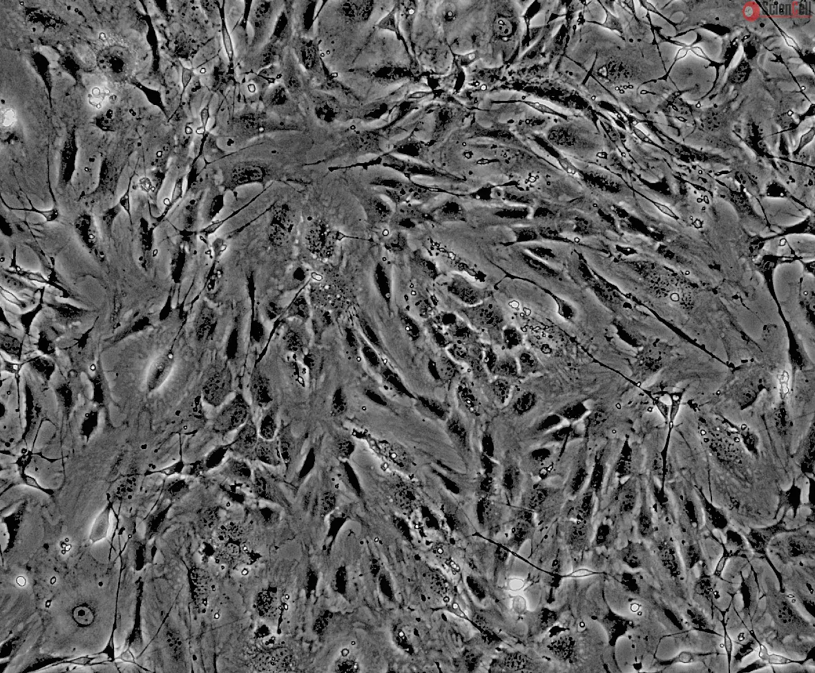
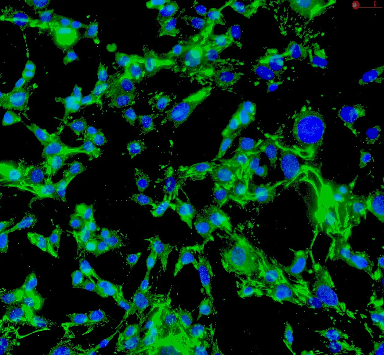
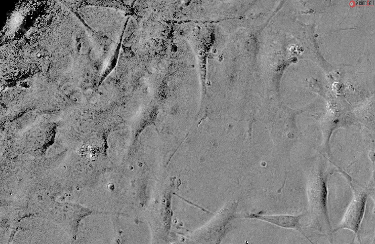
Mouse Perineurial Fibroblasts from CD1
Isolated from postnatal day 8 CD-1 mouse nerve. MPNF are cryopreserved at passage one and delivered frozen. Each vial contains >5 x 10⁵ cells in 1 ml volume.
Perineurial fibroblasts are of mesenchymal origin and form the perineurium. The perineurium plays an important role in maintaining the integrity of the internal peripheral nerve environment by creating a physical barrier that, under physiologic condition, limits the entry of biologically active proteins, infectious agents, and blood-borne cells into the nerve bundles. The perineurial fibroblasts are characterized by distinct ultrastructural features, including non-branching thin cytoplasmic processes coated by an external lamina and joined at their ends by a tight junction, few organelles, actin and vimentin filaments, and numerous pinocytotic vesicles. Perineurial fibroblasts are initially recruited from the surrounding mesenchyme to form a loose, permeable sheath around axons and Schwann cells, where they are separated by the extracellular matrix. These cells later undergo a mesenchymal-to-epithelial transition to form tight junctions and organize into the perineurium. Perineurial fibroblasts are immunoreactive for vimentin and epithelial membrane antigen but not for the Schwann cell marker S-100.
MPNF from ScienCell Research Laboratories are isolated from postnatal day 8 CD-1 mouse nerve. MPNF are cryopreserved at passage one and delivered frozen. Each vial contains >5 x 10⁵ cells in 1 ml volume. MPNF are characterized by immunofluorescence with antibodies specific to vimentin and/or fibronectin. MPNF are negative for mycoplasma, bacteria, yeast, and fungi. MPNF are guaranteed to further expand for 5 population doublings under the conditions provided by ScienCell Research Laboratories.
Recommended Medium
It is recommended to use Fibroblast Medium (FM, Cat. #2301) for culturing MPNF in vitro.