This study was performed to determine the association of Th17 cell phenotype with chronic
Renal proximal tubular epithelial cells play a central role in renal physiology and are among the cell types most sensitive to ischemia and xenobiotic nephrotoxicity. In ... More
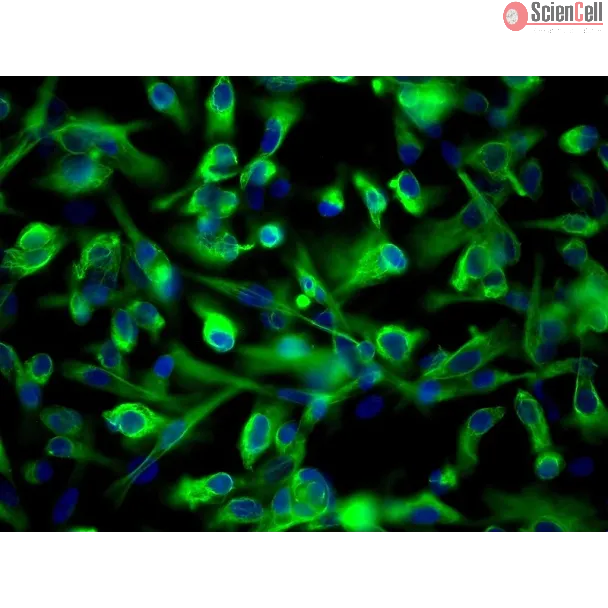
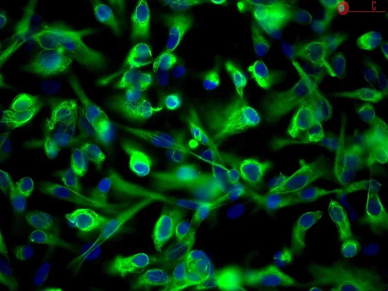
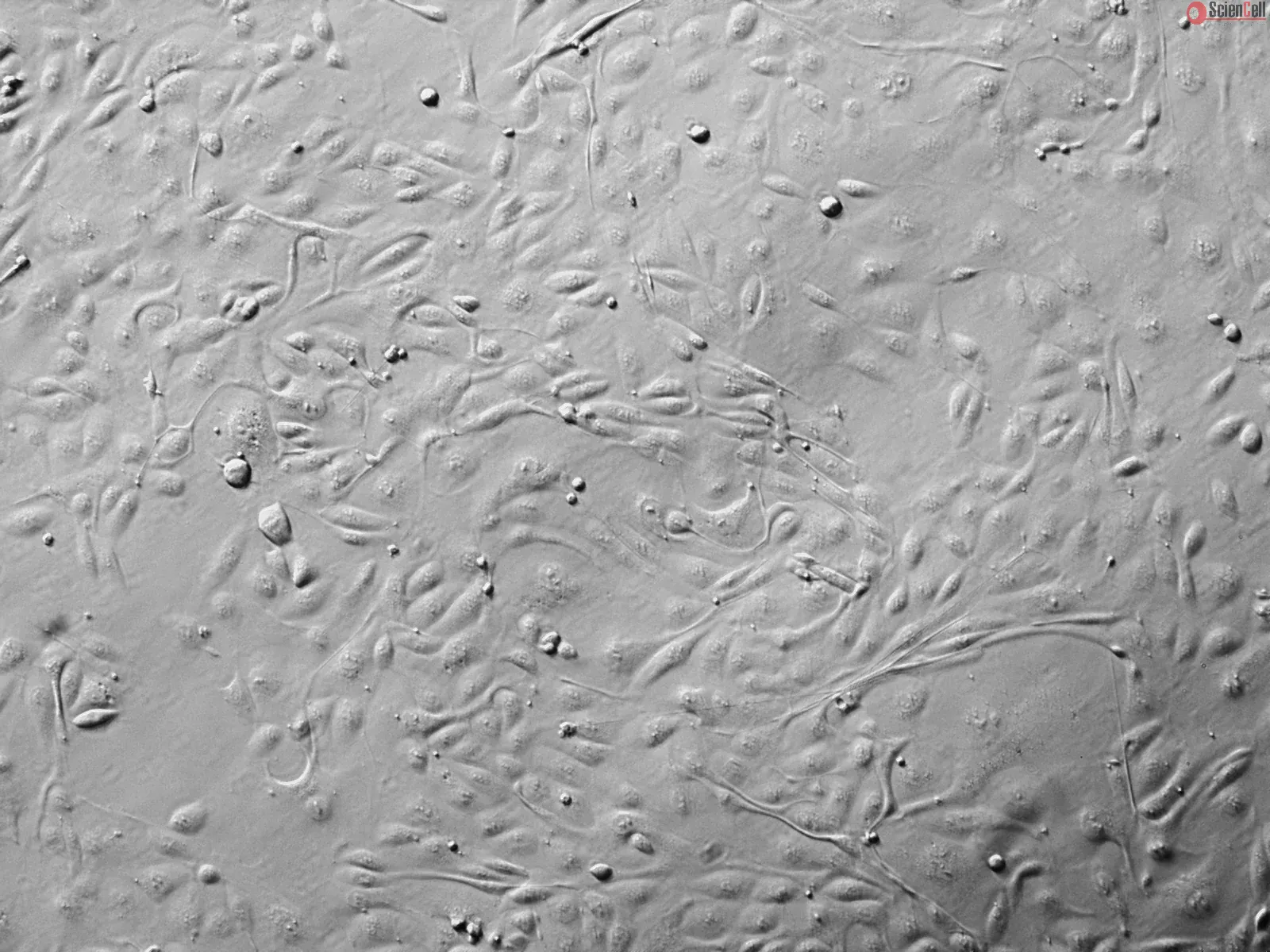
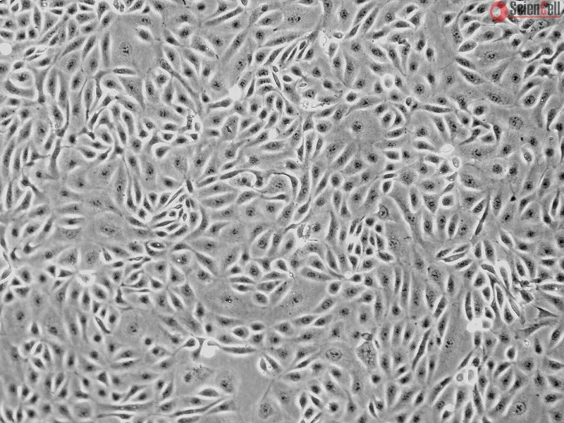
Renal proximal tubular epithelial cells play a central role in renal physiology and are among the cell types most sensitive to ischemia and xenobiotic nephrotoxicity. In order to investigate the molecular and cellular mechanisms underlying the pathophysiology of kidney injuries, a stable and well-characterized primary culture model of proximal tubular cells is required. An existing model of proximal tubular cells is hampered by the cellular heterogeneity of kidney; a method based on cell sorting for specific markers must therefore be developed. In this study, we present a primary culture model based on the mechanical and enzymatic dissociation of healthy tissue obtained from nephrectomy specimens. Renal epithelial cells were sorted using co-labeling for CD10 and CD13, two renal proximal tubular epithelial markers, by flow cytometry. Their purity, phenotypic stability and functional properties were evaluated over several passages. Our results demonstrate that CD10/CD13 double-positive cells constitute a pure, functional and stable proximal tubular epithelial cell population that displays proximal tubule markers and epithelial characteristics over the long term, whereas cells positive for either CD10 or CD13 alone appear to be heterogeneous. In conclusion, this study describes a method for establishing a robust renal proximal tubular epithelial cell model suitable for further experimentation. Less
Introduction: Based on some well-documented reports, we attempted to clarify the antifibrotic mechanisms of human Wharton's-jelly-derived mesenchymal stromal cells (WJ-MS... More
Introduction: Based on some well-documented reports, we attempted to clarify the antifibrotic mechanisms of human Wharton's-jelly-derived mesenchymal stromal cells (WJ-MSCs) from the perspective of induction of hepatocyte growth factor (HGF) expression in tubular epithelial cells (TECs). Less
Background/aims: Interleukin-17A (IL-17A) is a T cell-derived inflammatory cytokine that is upregulated during renal allograft rejection. The present study sought to furt... More
Background/aims: Interleukin-17A (IL-17A) is a T cell-derived inflammatory cytokine that is upregulated during renal allograft rejection. The present study sought to further describe the IL-17A-mediated proinflammatory/profibrotic activity of proximal tubule epithelium that may contribute to allograft rejection. Methods: Immortalized (HK-2) and primary (HRPTEpiC) human proximal tubule epithelial cells were utilized for this study. Profibrotic gene alterations were examined by real-time quantitative PCR. Inflammatory mediator secretion was examined by multiplex bead-based detection of secreted proteins. Immunofluorescence microscopy and immunoblotting were utilized to examine alterations in junctional protein expression and cell morphology. Results: In HK-2 cells IL-17A significantly downregulated the expression of the proepithelial gene CDH1 (E-cadherin) while the proinflammatory/profibrotic genes CTGF, CD44 and TGFBR1 were significantly increased. IL-17A also increased the secretion of fractalkine, G-CSF, GM-CSF, VEGF, IL-6 and IL-8. In HRPTEpiC 100 ng/ml IL-17A upregulated the proinflammatory/profibrotic genes ACTA2, CCL2, CHMP1A, CTGF, FN1, IL6, FSP1, SMAD1, SMAD5, TGFB1 and TGFBR2 while treatment with a reduced concentration of IL-17A (0.1 ng/ml) decreased SMAD5, TGFB1 and PDGFRB expression. Changes in ZO-1 and E-cadherin protein expression and cell morphology were examined following IL-17A treatment as indicators of epithelial-to-mesenchymal transition. IL-17A decreased ZO-1 expression in HK-2 and HRPTEpiC; however, E-cadherin was only reduced in HK-2 cells. Neither HK-2 nor HRPTEpiC assumed an elongated, fibroblast-like morphology following IL-17A treatment. Conclusions: IL-17A directly mediates proximal tubule epithelial cell proinflammatory/profibrotic activity as demonstrated by the alteration in genes associated with extracellular matrix remodeling and cell-cell interaction, and stimulation of inflammatory mediator and immune cell chemoattractant secretion. Additionally, IL-17A may have a negative impact on barrier integrity as indicated by ZO-1 downregulation. Copyright © 2010 S. Karger AG, Basel. Less
Extracellular matrix (ECM) coatings have been used to improve cell performance in bioartificial kidneys (BAKs). However, their effects on primary human renal proximal tub... More
Extracellular matrix (ECM) coatings have been used to improve cell performance in bioartificial kidneys (BAKs). However, their effects on primary human renal proximal tubule cells (HPTCs), which is the most important cell type with regard to clinical applications, have not been tested systematically. Also, the effects of ECM coatings on cell performance during extended time periods have not been addressed. Studying such effects is important for the development of long-term applications. Herein we analyzed for the first time systematically the effects of ECM coatings on proliferation and differentiation of human renal cells and we addressed, in particular, formation and long-term maintenance of differentiated epithelia. Our study focused on HPTCs. ECM coatings were tested alone or in combination with the growth factor bone morphogenetic protein-7 and other additives. The best results were obtained with ECMs consisting of the basal lamina components, laminin or collagen IV, and differentiated epithelia could be maintained up to three weeks on these ECMs. These results provide for the first time clear evidence which kinds of ECM coatings are most appropriate for BAKs. The results also showed that alpha-SMA-expressing myofibroblasts played a key role in the final disruption of differentiated epithelia. This suggests that epithelial-to-mesenchymal transition-related processes might be the major obstacle in long-term applications and such processes should be carefully addressed in future BAK-related research. Less
HIV-1 infection of renal cells has been proposed to play a role in HIV-1-associated nephropathy. Renal biopsy data further suggest that renal tubular cells may serve as r... More
HIV-1 infection of renal cells has been proposed to play a role in HIV-1-associated nephropathy. Renal biopsy data further suggest that renal tubular cells may serve as reservoir for HIV-1. The mechanism by which HIV-1 enters these cells has not been identified. Renal tubular cells do not express any of the known HIV-1 receptors, and our results confirmed lack of the expression of CD4, CCR5, CXCR4, DC-SIGN, or mannose receptors in tubular cells. The aim of this study, therefore, was to determine the mechanism that enables viral entry into renal tubular cells. An in vitro model was used to study the HIV-1 infection of human kidney tubular (HK2) cells and to identify the receptor that enables the virus to enter these cells. Results of these studies demonstrate that the C-type lectin DEC-205 acts as an HIV-1 receptor in HK2 cells. Interaction of HIV-1 with DEC-205 results in the internalization of the virus and establishment of a nonproductive infection. HIV-1-specific strong-stop DNA is detected in the infected HK2 cells for at least 7 d, and the virus can be transmitted in trans to sensitive target cells. HIV-1 entry is blocked by pretreatment with specific anti-DEC-205 antibody. Moreover, expression of DEC-205 in cells that lack the DEC-205 receptors renders them susceptible to HIV-1 infection. These findings suggest that DEC-205 acts as an HIV-1 receptor that mediates internalization of the virus into renal tubular cells, from which the virus can be rescued and disseminated by encountering immune cells. Less


,-1-mg-ml--2.jpg)