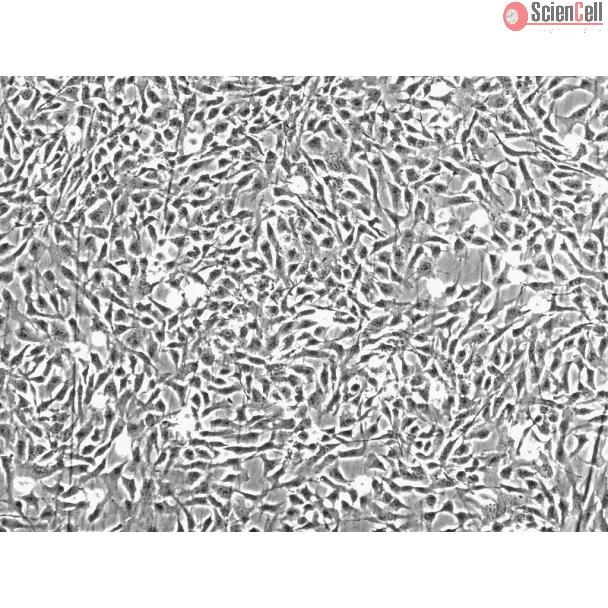
Chondrocytes are the resident cells of cartilage and are responsible for synthesizing a range of collagenous and non-collagenous extracellular matrix macromolecules. These include collagen type II, aggrecan, link protein, collagen type IX, and collagen type XI. The control of proliferation and differentiation of chondrogenic cells is central to the coordinated development of the vertebrate skeleton. Chondrocytes are capable of producing and responding to a large number of peptide growth factors and cytokines, including insulin-like growth factor-1 and interleukin-1. Chondrocyte cultures are useful in vitro models for studying cartilage regeneration and repair, cytokine and growth factor effects on cartilage, specific gene regulation and pathophysiology of arthritis.
HC-a from ScienCell Research Laboratories are isolated from human articular cartilage. HC-a are cryopreserved at passage one and delivered frozen. Each vial contains >5 x 10⁵ cells in 1 ml volume. HC-a are characterized by immunofluorescence with antibodies specific to S100B and/or type II collagen. HC-a are negative for HIV-1, HBV, HCV, mycoplasma, bacteria, yeast and fungi. HC-a are guaranteed to further expand for 15 population doublings under the conditions provided by ScienCell Research Laboratories.
Recommended Medium
It is recommended to use Chondrocyte Medium (CM, Cat. #4651) for the culturing of HC-a in vitro.
Osteoarthritis (OA) is characterized by degradation of the cartilage matrix, leading to pathologic changes in the joints. However, the pathogenic effects of synovial tiss... More
Osteoarthritis (OA) is characterized by degradation of the cartilage matrix, leading to pathologic changes in the joints. However, the pathogenic effects of synovial tissue inflammation on OA knees are not clear. To investigate whether the inflammation caused by the medial plica is involved in the pathogenesis of osteoarthritis, we examined the expression of matrix metalloproteinases (MMPs), tissue inhibitors of metalloproteinases (TIMPs), interleukin (IL)-1β, and tumor necrosis factor (TNF)-α in the medial plica and pannus-like tissue in the knees of patients with medial compartment OA who underwent either arthroscopic medial release (stage II; 15 knee joints from 15 patients) or total knee replacement (stage IV; 18 knee joints from 18 patients). MMP-2, MMP-3, MMP-9, IL-1β, and TNF-α mRNA and protein levels measured, respectively, by quantitative real-time PCR and Quantibody human MMP arrays, were highly expressed in extracts of medial plica and pannus-like tissue from stage IV knee joints. Immunohistochemical staining also demonstrated high expression of MMP-2, MMP-3, and MMP-9 in plica and pannus-like tissue of stage IV OA knees and not in normal cartilage. Some TIMP/MMP ratios decreased significantly in both medial plica and pannus-like tissue as disease progressed from stage II to stage IV. Furthermore, the migration of cells from the pannus-like tissue was enhanced by IL-1β, while plica cell migration was enhanced by TNF-α. The results suggest that medial plica and pannus-like tissue may be involved in the process of cartilage degradation in medial compartment OA of the knee. Less
Background In addition to treating acute promyelocytic leukemia, arsenic trioxide (ATO) suppresses other solid tumors, including chondrosarcoma. However, the effects of A... More
Background In addition to treating acute promyelocytic leukemia, arsenic trioxide (ATO) suppresses other solid tumors, including chondrosarcoma. However, the effects of ATO on metastasis in chondrosarcoma cells, and the underlying molecular mechanisms remain unclear. Methods The effects of ATO on the migratory and invasive capacities of chondrosarcoma cells were investigated by Wound healing, Transwell and EMT assays. The expression of miR-125b in human chondrosarcoma tissues and cell lines was detected by real-time PCR analysis. Bisulfite sequencing analysis (BSP) was used to detect the effects of ATO on the expression of miR-125b. The gain-of-function and loss-of-function experiments were performed on chondrosarcoma cell lines to investigate the effects of miR-125b on chondrosarcoma invasion, and to determine whether signal transducer and activator of transcription 3(Stat3) mediates these effects. Dual-luciferase reporter assay was used to identify whether Stat3 is a direct target of miR-125b. Results MiR-125b was significantly downregulated in human metastatic chondrosarcoma tissues and cell lines but not in non-metastatic chondrosarcoma tissues. ATO up-regulates the expression of miR-125b by the demethylation of DNA. ATO induces MET and attenuates the invasive capacities of chondrosarcoma cells through miR-125b. Stat3 was verified as a direct target of miR-125b, which is involved in ATO regulating EMT-associated traits. Conclusions These findings, for the first time, provides evidence that the miR-125b-mediated inhibition of Stat3 is involved in the ATO-induced attenuation of metastasis in chondrosarcoma cells. Less
ScienCell Research Laboratories (SRL) takes pride in being a resource for researchers all over the world. The publications listed here are not meant as an endorsement or confirmation of the reliability of the products.


,-1-mg-ml--2.jpg)