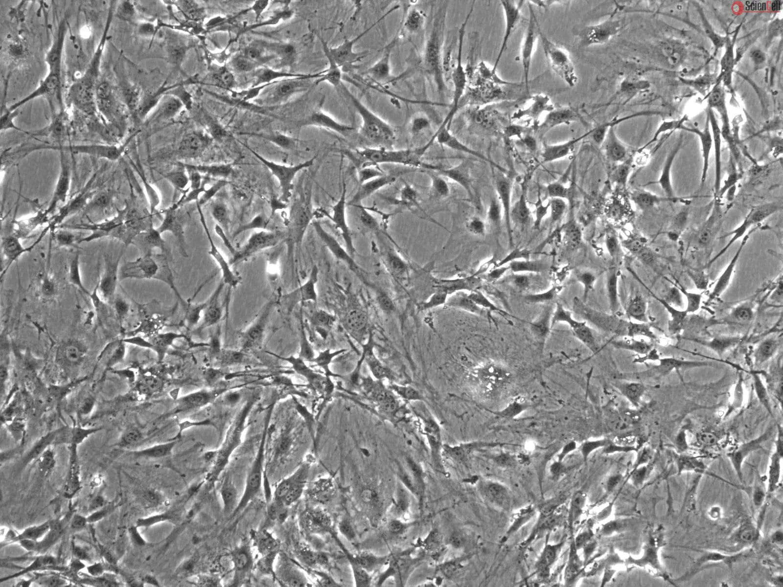
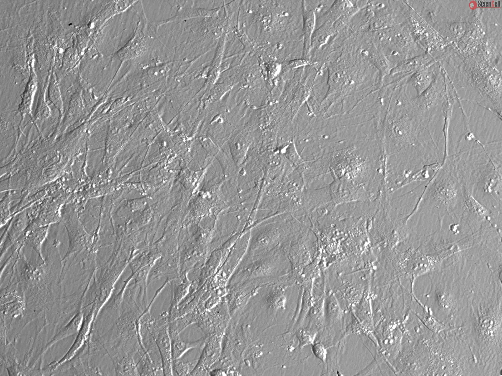
Brain Vascular Pericytes (BVP) are perivascular cells that are closely associated with the endothelium of capillaries and other small vessels. BVP, located between endothelial cells and astrocytes in the brain, communicate with other cells by extending long cytoplasmic processes which wrap around the capillaries. BVP participate in a variety of processes including angiogenesis, endothelial cell survival, regulation of capillary blood flow, and establishment and maintenance of the blood-brain barrier. Pericyte dysregulation has been linked to several pathological conditions such as hypertension, diabetic retinopathy, atherosclerosis, multiple sclerosis, Alzheimer’s disease, and tumor angiogenesis. The unique and diverse functions of BVP make them novel candidates for cell therapy in regenerative medicine. Cultured primary mouse BVP (MBVP) are a useful in vitro model for understanding the molecular mechanisms of blood-brain barrier regulation and for studying a wide variety of central nervous system diseases.
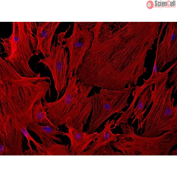
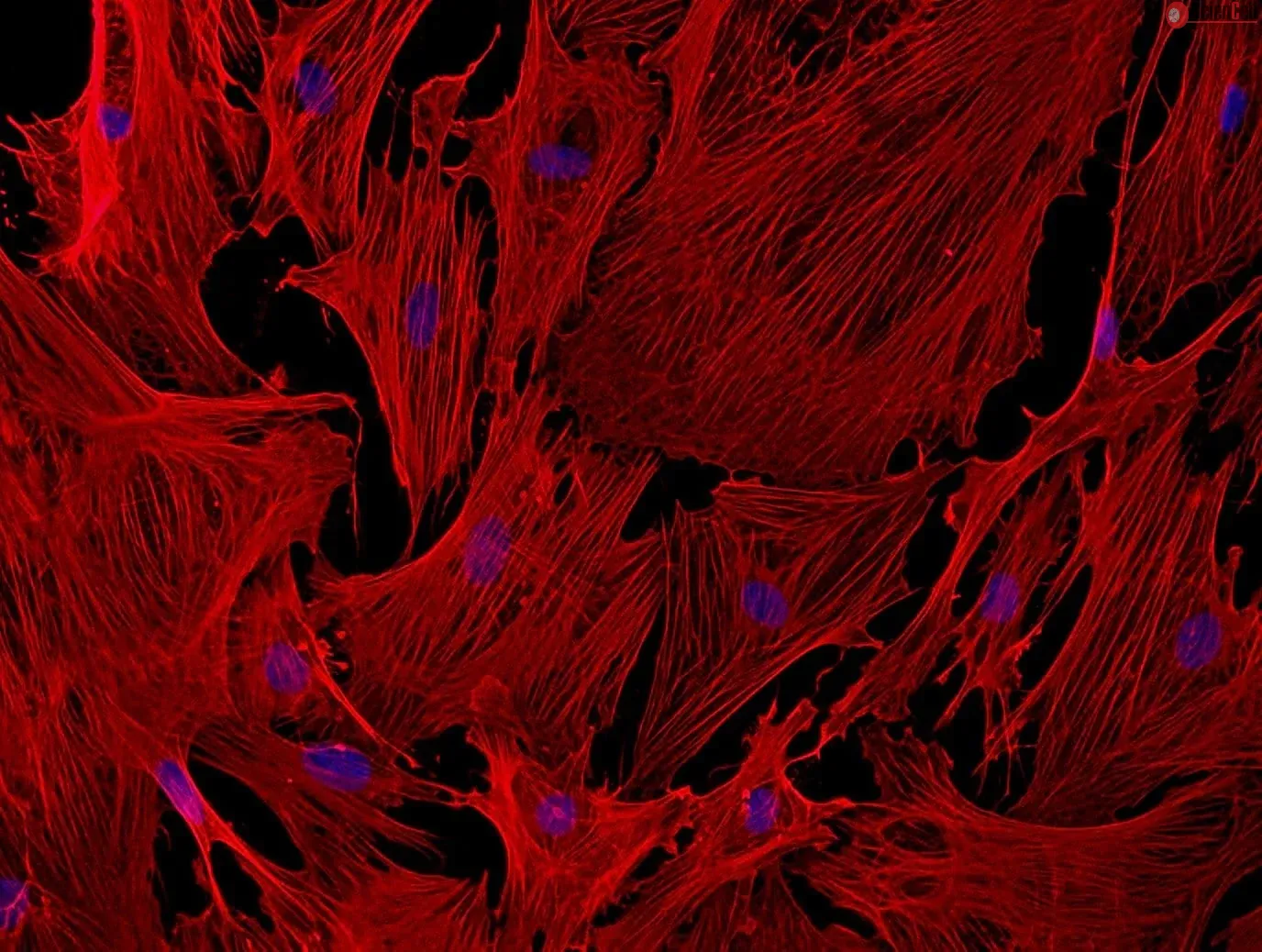
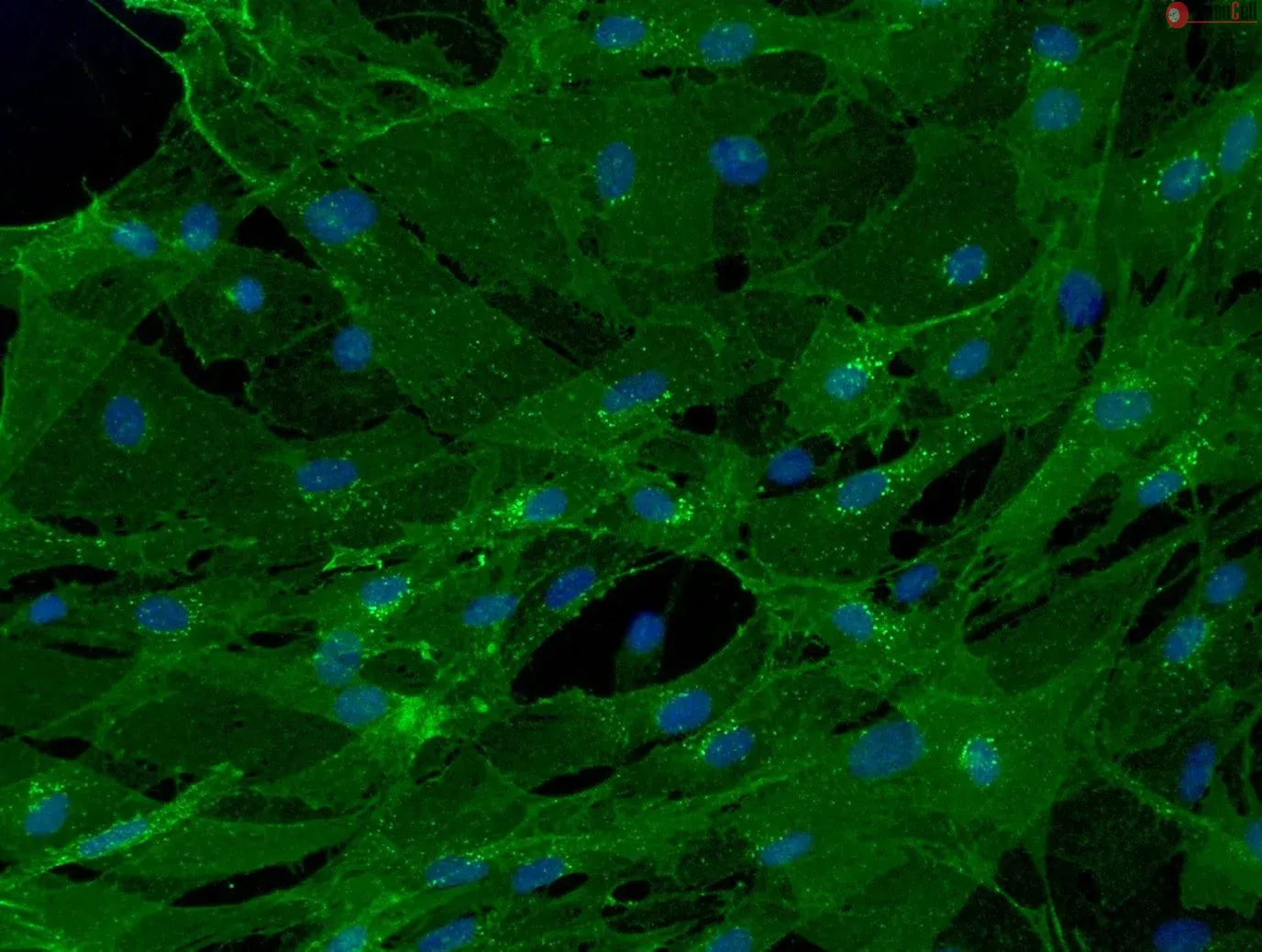
MBVP from ScienCell Research Laboratories are isolated from adult C57BL/6 mouse brain. MBVP are cryopreserved at passage one and delivered frozen. Each vial contains >5 x 10^5 cells in 1 ml volume. MBVP are characterized by immunofluorescence with antibody specific to platelet-derived growth factor-β ( PDGF Receptor-β) and α-smooth muscle actin. MBVP are negative for mycoplasma, bacteria, yeast, and fungi. MBVP are guaranteed to further expand for 5 population doublings under the conditions provided by ScienCell Research Laboratories.
Recommended Medium
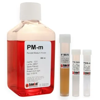
It is recommended to use Pericyte Medium-mouse (PM-m, Cat. #1231) for culturing MBVP in vitro.
Product Sheets
 M1200-57
Search for a Certificate of Analysis by Lot Number below:
Background
While the role of pericytes in blood-brain barrier (BBB) disruption and neuroinflammation is well-established in adult neurological disorders, their contribut... More
Background
While the role of pericytes in blood-brain barrier (BBB) disruption and neuroinflammation is well-established in adult neurological disorders, their contribution to neonatal brain injury is largely unexplored. Here, we investigated the role of brain pericytes in hypoxic-ischemic (HI) brain injury in the developing brain, with a particular focus on the regulatory role of pericyte-derived microRNA210 (miR210) in pericyte dysfunction.
Methods
HI brain injury was induced on postnatal day 9 transgenic mice, including Atp13a5-tdTomato brain pericyte reporter mice, pericyte-specific diphtheria toxin receptor mice, miR210 knockout mice, and wild-type controls. Post-injury assessments include brain infarct, brain edema, BBB permeability, ELISA, western blotting, immunostaining, and neurological function test. BBB-associated cells, including pericytes and endothelial cells, were isolated from mouse brain using an immunomagnetic approach. RNA sequencing analysis was conducted to examine transcriptomic changes in pericytes after HI. To investigate the regulatory role of miR210 in pericyte dysfunction and its underlying mechanisms, primary pericytes were transfected with miR210 mimic or negative control, followed by oxygen-glucose deprivation. Transfected cells were also treated with either interleukin 1 type 1 receptor neutralizing antibody or recombinant interleukin 1 type 2 receptor chimera protein. Post-assays included RT-qPCR, immunostaining and cell viability assay. Student’s t test or one-way ANOVA followed by Bonferroni test was used, as appropriate.
Results
HI resulted in a time-dependent loss of pericytes in pericyte reporter mouse pups. Ablation of brain pericytes exacerbated BBB disruption and HI brain injury in neonatal brain. miR210 deletion mitigated brain pericyte loss and BBB leakage post-HI. Transcriptomic analysis revealed that HI-induced pericyte dysfunction was associated with upregulated genes enriched in biological processes such as “cellular response to interleukin 1”. miR210 knockout suppressed the expression of proinflammatory markers such as Il1r1. Mechanistically, miR210 overexpression increased proinflammatory cytokine levels and promoted pericyte cell death under oxygen-glucose deprivation, effects that were reversed by IL1R1 blockade. Importantly, brain pericyte-specific miR210 deletion preserved pericyte viability and BBB integrity, and provided neuroprotection after HI.
Conclusions
These findings underscore the critical role of brain pericytes in BBB function in the developing brain and identify miR210 as a central regulator of pericyte dysfunction following neonatal HI brain injury. Less
Pericytes line the microvasculature throughout the body and play a key role in regulating blood flow by constricting and dilating vessels. However, the biophysical mechan... More
Pericytes line the microvasculature throughout the body and play a key role in regulating blood flow by constricting and dilating vessels. However, the biophysical mechanisms through which pericytes transduce microenvironmental chemical and mechanical cues to mediate vessel diameter, thereby impacting oxygen and nutrient delivery, remain largely unknown. This knowledge gap is clinically relevant as numerous diseases are associated with the aberrant contraction of pericytes, which are unusually susceptible to injury. Here, we report the development of a high-throughput hydrogel-based pericyte contraction cytometer that quantifies single-cell contraction forces from murine and human pericytes in different microvascular microenvironments and in the presence of competing vasoconstricting and vasodilating stimuli. We further show that murine pericyte survival in hypoxia is mediated by the mechanical microenvironment and that, paradoxically, pre-treating pericytes to reduce contraction increases hypoxic cell death. Moreover, using the contraction cytometer as a drug-screening tool, we found that cofilin-1 could be applied extracellularly to release murine pericytes from hypoxia-induced contractile rigor mortis and, therefore, may represent a novel approach for mitigating the long-lasting decrease in blood flow that occurs after hypoxic injury. Less
ScienCell Research Laboratories (SRL) takes pride in being a resource for researchers all over the world. The publications listed here are not meant as an endorsement or confirmation of the reliability of the products.